
Whole Spine MRI
The human spine is a complex structure consisting of bones, discs, nerves, and muscles that support the body’s weight and facilitate movement. An MRI (Magnetic Resonance Imaging) scan of the whole spine is a non-invasive imaging technique used to visualise detailed structures of the spine. This advanced diagnostic tool helps in the assessment and diagnosis of various spinal conditions, providing critical information that aids in effective treatment planning. Book MRI Spine in Barast for7000*
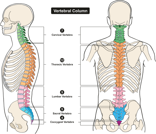
Anatomy Of The Spine
Cervical Region: Encompasses the neck and comprises 7 cervical vertebrae.
Thoracic Region: Located in the chest area, it consists of 12 thoracic vertebrae.
Lumbar Region: Situated in the lower back, it comprises 5 lumbar vertebrae, which are the largest and strongest.
Sacrum: The sacrum is a triangular bone formed by the fusion of 5 sacral vertebrae. It connects the spine to the pelvis.
Coccyx: Commonly known as the tailbone, the coccyx consists of 4 fused coccygeal vertebrae.
MRI Whole Spine Available In
Name
Proc. Time
Rating
Price

What Is A Spine MRI?
A Whole Spine MRI involves using magnetic fields and radio waves to produce detailed images of the cervical, thoracic, and lumbar regions of the spine. Unlike X-rays or CT scans, MRI does not use ionising radiation, making it a safer option for detailed imaging. The procedure typically takes 45 minutes to an hour and can capture images in multiple planes, offering a comprehensive view of the spinal anatomy.
What Is Its Purpose?
An MRI scan of the whole spine is a crucial diagnostic tool that provides detailed images of spinal structures, aiding in the diagnosis and management of various conditions. It helps identify spinal disorders such as herniated discs, degenerative disc disease, spinal stenosis, and spondylolisthesis. The MRI detects tumours and lesions, assessing their size, location, and extent, which is essential for conditions like multiple sclerosis and spinal cancers.
The scan provides insights into the vertebral bodies, revealing abnormalities in structure, alignment, or integrity such as fractures, deformities, or osteoporosis. It captures the state of intervertebral discs, showcasing signs of degeneration like disc desiccation or herniation, which can contribute to symptoms such as back pain or nerve compression. Additionally, the MRI visualises the spinal cord and nerve roots, detecting lesions, inflammation, or injuries that may cause neurological symptoms like numbness, weakness, or radiculopathy. It evaluates the spinal canal for stenosis or the presence of tumours, which can lead to compression of neural structures. Furthermore, the MRI assesses facet joints, ligaments, soft tissues, and bone marrow, identifying degenerative changes, injuries, or neoplasms that may affect spinal health. It highlights vascular abnormalities and provides enhanced visualisation of vascular structures with contrast agents. Book MRI Spine in Barast for7000*
Types Of Spine MRIs And Their Role In Early Disease Detection
Cervical Spine MRI: A Cervical Spine MRI scans the neck and upper back, focusing on the C1-C7 vertebrae. This MRI examines the soft tissues, cervical vertebrae, intervertebral discs, and spinal cord. It is useful for detecting issues such as herniated discs, spinal stenosis, tumours, and multiple sclerosis in the neck region. By identifying nerve compression and sources of neck pain or radiculopathy, this MRI facilitates timely and effective treatment.
Thoracic Spine MRI: A Thoracic Spine MRI targets the mid-back, focusing on the T1-T12 vertebrae. It captures detailed images of the thoracic vertebrae, spinal cord, and related structures. This type of MRI is essential for diagnosing herniated discs, spinal tumours, and degenerative diseases in the mid-back. It also identifies fractures, infections, and inflammatory conditions. Since the thoracic spine affects the function of the hands, arms, chest, and abdominal muscles, early detection through MRI helps in managing conditions that impair these functions.
Lumbar Spine MRI: A Lumbar Spine MRI scans the lower back, focusing on the L1-L5 vertebrae. These vertebrae support a significant amount of body weight and absorb more shock than other parts of the spine. This MRI is crucial for diagnosing conditions such as degenerative disc disease, herniated discs, lumbar stenosis, and sciatica. By pinpointing the cause of lower back pain and leg pain early, it prevents the progression of spinal issues.
Whole Spine MRI: A Whole Spine MRI provides a comprehensive view of the entire spinal column, including the cervical, thoracic, and lumbar regions. It is particularly useful for evaluating widespread/complex spinal conditions, such as metastatic cancer, multiple sclerosis, or diffuse spinal infections. This comprehensive imaging is beneficial for patients with symptoms affecting multiple regions of the spine, ensuring thorough diagnosis and management.
Understand How These MRIs Help In Early Detection Of Diseases
These MRI scans offer high-resolution images that reveal even small abnormalities in the spine’s structure, facilitating early detection before issues become severe. The non-invasive nature of MRI makes it suitable for regular monitoring, and the use of contrast agents can enhance the visibility of certain abnormalities, aiding early diagnosis of tumours, infections, and inflammatory conditions. Multiplanar imaging captures images in multiple planes, offering a comprehensive view, and the superior soft tissue contrast of MRI helps in detecting issues related to intervertebral discs, spinal cord, and nerve roots. By utilising these different types of spine MRI, healthcare providers can accurately diagnose and treat a variety of spinal conditions early, improving patient outcomes and preventing the progression of spinal diseases. Book MRI Spine in Barast for7000*
Diseases Diagnosed By Whole Spine MRI
A whole spine MRI is an invaluable diagnostic tool that helps in the identification & evaluation of a wide range of spinal diseases and conditions. Here are the diseases that can be diagnosed via this advanced imaging technique:
Degenerative Disc Disease: Degenerative disc disease occurs when the intervertebral discs, which act as cushions between the vertebrae, begin to deteriorate. MRI can detect changes in the discs, such as dehydration, bulging, and herniation, which are indicative of this condition. The causes include aging, genetics, stress, injury, obesity, smoking, and poor posture.
Herniated Discs: A herniated/slipped/ruptured disc occurs when the soft inner core of the disc protrudes through a tear in the outer layer. MRI provides images of disc herniations and the extent to which surrounding nerves are compressed.
Spinal Stenosis: Spinal stenosis is the narrowing of the spinal canal, which can put pressure on the spinal cord and nerves. MRI can identify the exact locations and severity of the narrowing, as well as its impact on nerve tissues.
Spinal Tumours: MRI is highly effective in detecting tumours within the spine, including benign and malignant growths. It helps in determining the size, location, and extent of the tumours, as well as their relation to surrounding tissues.
Infections: Spinal infections, such as osteomyelitis (infection of the bone) or discitis (infection of the intervertebral disc), can be detected through MRI. The scan can show inflammatory changes, abscesses, and other signs of infection.
Multiple Sclerosis (MS): MRI is a crucial tool in the diagnosis and monitoring of multiple sclerosis. It can reveal plaques or lesions in the spinal cord that are characteristic of MS, helping to assess the extent and activity of the disease.
Spinal Cord Injuries: MRI provides detailed images of the spinal cord and surrounding structures, making it ideal for evaluating spinal cord injuries. It can detect damage from trauma, including bruising, bleeding, or severing of the spinal cord.
Spondylolisthesis: This condition occurs when a vertebra slips out of place onto the bone below it. MRI can help determine the degree of slippage and its effect on the spinal cord and nerves.
Arthritis: Various forms of arthritis, such as osteoarthritis and rheumatoid arthritis, can affect the spine. MRI can show joint damage, inflammation, and other changes associated with these conditions.
Congenital Spine Disorders: MRI can identify congenital anomalies of the spine, such as scoliosis (curvature of the spine), kyphosis (excessive outward curve), and other developmental abnormalities.
Vascular Abnormalities: MRI can detect vascular abnormalities in the spine, such as arteriovenous malformations (AVMs), which are abnormal tangles of blood vessels that can cause pain and other symptoms.
Who Should Get Tested?
MRI of the whole spine may be recommended for individuals who present with symptoms or conditions that warrant further evaluation of spinal structures. Here are people who should book a whole spine MRI:
Individuals With Persistent/Severe Back Pain: Individuals experiencing persistent or severe back pain that does not improve with conservative treatments may benefit from a whole spine MRI to identify potential causes such as disc herniation, spinal stenosis, or degenerative disc disease.
Patients With Neck Pain & Radicular Symptoms: Patients with neck pain accompanied by radiating symptoms such as arm pain, numbness, or weakness may require a whole spine MRI to assess cervical spine structures and identify potential nerve compression or other issues.
Individuals With Neurological Symptoms: Individuals presenting with neurological symptoms such as numbness, tingling, weakness, or loss of coordination in the arms or legs may need a whole spine MRI to evaluate spinal cord and nerve root involvement.
Individuals With Suspected Disc Herniation/Degeneration: Patients with symptoms suggestive of disc herniation, such as shooting pain or sciatica, or those with suspected disc degeneration may be candidates for a whole spine MRI to visualise the affected discs and assess their condition.
Individuals With Traumatic Spinal Injuries: Individuals who have experienced a traumatic injury to the spine, such as a car accident or fall, may require a whole spine MRI to assess for fractures, ligament injuries, spinal cord damage, or soft tissue trauma.
Patients With Chronic Conditions: Patients with chronic conditions affecting the spine, such as multiple sclerosis or rheumatoid arthritis, may undergo periodic whole spine MRIs to monitor disease progression and assess treatment efficacy.
Individuals Undergoing Pre-Surgical Planning & Post-Surgical Assessment: Individuals scheduled for spinal surgery may undergo a whole spine MRI for pre-surgical planning to evaluate the extent of spinal pathology and identify any abnormalities. Similarly, post-surgical assessment with a whole spine MRI may be necessary to evaluate surgical outcomes and detect any post-operative complications.
It’s important to note that the decision to undergo a whole spine MRI should be made in consultation with a healthcare provider, who can assess individual symptoms, medical history, and clinical indications to determine the necessity of the imaging test. Book MRI Spine in Barast for7000*
MRI Whole Spine Available In
| MRI Scans | City | Price | ||
|---|---|---|---|---|
| 1 | MRI Whole Spine | - | 7000 |
How Does A Whole Spine MRI Work?
A whole spine MRI, or magnetic resonance imaging, works by utilising powerful magnets and radio waves to create detailed images of the entire spinal column, from the cervical to the coccygeal regions.
Magnetic Field Generation: The MRI machine generates a strong magnetic field around the patient’s body. This magnetic field aligns the hydrogen atoms in the body’s tissues, particularly in water molecules.
Radiofrequency Pulse: Short bursts of radiofrequency pulses are then directed at the aligned hydrogen atoms. These pulses temporarily disrupt the alignment of the hydrogen atoms.
Return to Alignment: After the radiofrequency pulse is turned off, the hydrogen atoms return to their original alignment with the magnetic field. As they do so, they emit energy, which is detected by the MRI machine.
Signal Detection: The emitted energy is captured by specialised coils placed around the area of interest, in this case, the entire spine. These coils act as receivers and transmit the energy signals to a computer.
Image Reconstruction: The computer processes signals received from the coils to create cross-sectional images of the spinal structures. Different imaging sequences, such as T1-weighted, T2-weighted, and STIR (Short Tau Inversion Recovery) sequences, provide unique information about the spinal anatomy & pathology.
Multiplanar Imaging: The MRI machine can acquire images in multiple planes, including sagittal (side-to-side), axial (horizontal), and coronal (front-to-back) planes. This allows for a comprehensive evaluation of the entire spine from various perspectives.
Contrast Enhancement (Optional): In some cases, a contrast agent, typically containing gadolinium, may be injected intravenously to enhance the visibility of certain structures or abnormalities. This contrast agent can help highlight areas of inflammation, tumours, or vascular abnormalities.
Scan Completion: Once all necessary imaging sequences have been acquired, the MRI scan is complete. The images are reviewed by a radiologist, who interprets the findings and generates a report for the physician.
How Is This Scan Done?
A Spine MRI scan is performed in a methodical way to ensure detailed and accurate images of the spinal column.
Preparation
Remove Metal Objects: The patient is asked to remove all metal objects, such as jewellery, watches, hairpins, and eyeglasses. This is crucial as metal can interfere with the MRI’s magnetic field.
Medications: The patient should inform the technologist about any medications they are taking and follow specific instructions regarding any medications that may need to be adjusted before the scan.
Claustrophobia: If the patient has a fear of enclosed spaces, they should discuss this with their healthcare provider. Strategies such as medication or the use of an open MRI machine may be recommended.
Positioning
Lying on the Table: The patient lies down on the MRI table, usually in a supine (face-up) position. The head is positioned in a headrest, and the body is aligned with the bore (opening) of the MRI machine.
Coil Placement: MRI coils are placed around the regions of the spine to be imaged. These coils act as antennas to capture the MRI signals.
Entering the MRI Machine
Moving into the Bore: The MRI table slowly moves into the bore of the MRI machine. The part of the spine to be scanned is positioned in the centre of the magnet.
Communication: Throughout the scan, the technologist communicates with the patient via an intercom system. The patient may also be given a call button to alert the technologist if they need assistance.
Scan Acquisition
Magnetic Field and Radio Waves: Inside the MRI machine, the patient is exposed to a strong magnetic field and radiofrequency pulses. These interact with the hydrogen atoms in the body to create detailed images.
Image Sequences: Multiple imaging sequences are performed to capture different types of images, such as T1-weighted, T2-weighted, and STIR sequences. Each sequence provides unique information on the spinal structures.
Contrast Dye: In some cases, the patient may need a contrast dye injection to enhance the visibility of certain structures. If contrast dye is used, it will be administered midway through the scan. The patient should disclose any kidney issues or allergies to the dye before the procedure.
Motion Control
Remaining Still: To obtain clear images, it’s crucial for the patient to remain still during the scan. Even slight movements can result in motion artefacts that degrade image quality.
Breath-Holding: Patients may be asked to hold their breath briefly during certain sequences to minimise motion.
Post-Scan
Completion: Once the scan is complete, the patient is slowly moved out of the MRI machine.
Recovery: Patients can resume their normal activities immediately after the scan. There is no recovery time or side effects associated with MRI.
Image Analysis
Radiologist Review: The MRI images are analysed by a radiologist, who interprets them and generates a report.
Results Discussion: The results are typically discussed with the patient by their referring physician, who may recommend further testing or treatment based on the findings.
The duration of an MRI of the whole spine can vary depending on several factors, including the specific protocol used by the imaging facility, the patient's individual anatomy, and whether contrast agent administration is required. However, on average, a whole spine MRI typically takes between 45 minutes to 1 hour to complete. This timeframe includes the time needed for patient preparation, positioning on the MRI table, image acquisition, and any necessary adjustments during the scan. It’s essential to remain as still as possible during the procedure to ensure clear and accurate imaging results. Book MRI Spine in Barast for7000*
NORMAL RESULTS
A normal MRI of the whole spine typically indicates that there are no significant abnormalities or pathologies detected in the various structures of the spine. This includes the vertebrae, intervertebral discs, spinal cord, nerve roots, ligaments, and surrounding soft tissues. Let’s understand the result in detail:
Vertebrae: The vertebrae appear normal in shape, size, and alignment, without any evidence of fractures, tumours, or other abnormalities.
Intervertebral Discs: The intervertebral discs exhibit a normal signal intensity and height, indicating adequate hydration and no signs of degeneration, herniation, or bulging.
Spinal Cord and Nerve Roots: The spinal cord and nerve roots appear unremarkable, without any signs of compression, inflammation, or abnormal enhancement.
Soft Tissues and Ligaments: The soft tissues and ligaments surrounding the spine appear intact, without any evidence of injury, inflammation, or masses.
No Other Pathologies: No other abnormalities such as cysts, tumours, infections, or vascular malformations.
ABNORMAL RESULTS
An abnormal MRI of the whole spine indicates the presence of one or more abnormalities or pathologies within the various structures of the spine. These abnormalities may range in severity and can involve different parts of the spine, including the vertebrae, intervertebral discs, spinal cord, nerve roots, ligaments, and surrounding soft tissues. Here’s what an abnormal MRI of the whole spine may indicate:
Vertebral Abnormalities: This may include fractures, deformities, tumours, infections (such as osteomyelitis), or degenerative changes (such as osteoarthritis or spondylolisthesis).
Intervertebral Disc Abnormalities: Abnormalities of the intervertebral discs may include degeneration, herniation, bulging, or inflammation. These can lead to compression of nearby structures, such as nerve roots or the spinal cord, causing symptoms such as pain, numbness, or weakness.
Spinal Cord or Nerve Root Compression: Compression of the spinal cord or nerve roots may occur due to disc herniation, spinal stenosis (narrowing of the spinal canal), tumours, cysts, or other structural abnormalities. This can result in symptoms such as radiculopathy (radiating pain, numbness, or weakness) or myelopathy (weakness or paralysis).
Soft Tissue Abnormalities: Abnormalities of the ligaments, muscles, or other soft tissues surrounding the spine may indicate injury, inflammation, infection, or tumours.
Other Pathologies: Abnormalities such as spinal tumours (benign or malignant), cysts, vascular malformations, or infections may also be detected on an abnormal MRI of the whole spine.
Overall, an abnormal MRI of the spine suggests the presence of underlying spinal pathology that may require further evaluation and treatment. It’s essential for the physician to correlate these findings with the patient’s clinical history and symptoms to guide appropriate care. Book MRI Spine in Barast for7000*
Associated Risks
Magnetic Field Exposure: The MRI’s strong magnetic field can interfere with or damage implanted devices like pacemakers and can cause metal in the body to heat up or move. Inform your provider of any implants or metal fragments.
Contrast Agents: Gadolinium, used to enhance MRI images, can cause allergic reactions or, rarely, nephrogenic systemic fibrosis (NSF) in individuals with impaired kidney function. Always inform your provider of any kidney issues or allergies.
Claustrophobia: The enclosed space of the MRI machine can cause anxiety. Discuss any concerns with your provider beforehand, who may offer sedation or relaxation techniques.
Noise: The loud noises during the scan can be uncomfortable, but earplugs or headphones are usually provided to reduce noise exposure.
Motion Artefacts: Movement during the scan can blur images, requiring repeat scans. Patients must remain still to ensure clear images.
Pregnancy: Non-urgent MRIs are generally avoided during the first trimester. If a contrast agent is needed, the risks and benefits should be carefully evaluated.
Temporary Discomfort: Lying still on the hard MRI table can cause discomfort, especially for those with back pain. Communicate any discomfort to the technologist for adjustments.
Precautions To Avoid Spine-Related Diseases
Maintaining spinal health is crucial for avoiding spine-related diseases. Here are some essential precautions:
Maintain Good Posture: The patient should stand and sit correctly, ensuring the back is straight and the shoulders are aligned. When sitting, the feet should be flat on the floor, and slouching should be avoided to reduce spinal strain.
Exercise Regularly: Regular exercise strengthens core muscles, which support the spine. Activities like yoga, pilates, and core workouts are beneficial. They ensure a healthy weight, reducing pressure on the spine.
Lift Properly: The patient should use their legs rather than their back when lifting heavy objects. Bending at the knees and keeping the object close to the body can prevent back injuries.
Maintain A Healthy Weight: Managing weight through a balanced diet and regular exercise is important. Excess weight, particularly around the abdomen, can strain the lower back.
Stay Hydrated: Drinking plenty of water is essential for the health of intervertebral discs, which have a high water content. Proper hydration helps maintain disc flexibility and health.
Use Ergonomic Furniture: Utilising chairs and mattresses that provide good support helps maintain the natural curve of the spine. This can prevent back pain and promote overall spinal health.
Avoid Prolonged Sitting: Taking regular breaks to stand, stretch, and walk can reduce stress on the spine. This is especially important for individuals with sedentary jobs.
Wear Proper Footwear: Wearing shoes that offer good support and cushioning is important for maintaining proper spine alignment and reducing the risk of back pain.
Quit Smoking: Smoking reduces blood flow to the spine, which can contribute to disc degeneration. Quitting smoking improves overall health and spinal health.
Regular Check-Ups: The patient should visit healthcare providers regularly for check-ups. Early detection of spine-related issues can prevent more serious problems. Persistent back pain or other symptoms should be promptly addressed by a professional.