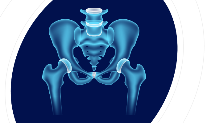
What Is A Pelvis CT Scan?
A computed tomography (CT) scan of the pelvis is a non-invasive diagnostic imaging test that uses x-rays to create detailed cross-sectional images of the pelvic region. This includes vital organs such as the bladder, reproductive organs, parts of the intestines, and major blood vessels. Unlike regular x-rays, which provide limited information, a CT scan combines multiple x-ray images taken from different angles and uses computer processing to produce comprehensive, high-resolution pictures of the internal structures. This enhanced imaging capability allows doctors to diagnose, evaluate, and monitor a variety of conditions and diseases in the pelvic area with greater accuracy.
Pelvis CT Scan Purpose
The primary purpose of a pelvis CT scan is to obtain detailed, cross-sectional images of the pelvic region, which includes vital organs such as the bladder, reproductive organs, and portions of the intestines. This imaging test is instrumental in diagnosing unexplained abdominal and pelvic pain by revealing underlying conditions like infections, inflammations, or structural abnormalities. It plays a crucial role in detecting and evaluating tumours, cysts, and other abnormal masses, providing essential information on their size, location, and extent, which is vital for treatment planning. Additionally, a pelvis CT scan is invaluable in assessing trauma-related injuries, such as fractures or internal bleeding, following accidents or falls. It guides surgical procedures by offering a clear view of the pelvic anatomy, thereby minimising risks and improving outcomes. Moreover, it helps monitor disease progression and treatment response in patients with chronic conditions like cancer or inflammatory diseases. The scan can also identify infections and inflammations such as appendicitis or pelvic inflammatory disease, aiding in timely and accurate diagnosis. When performed with contrast dye, it highlights blood vessels, assisting in the detection of vascular issues like blood clots or aneurysms. Overall, the pelvis CT scan is a versatile and critical tool in the comprehensive evaluation of pelvic health concerns. Book a PELVIS CT SCAN in Chaksu for ONLY5000*
NCCT Pelvis Available In
Name
Proc. Time
Rating
Price


What Does A Pelvis CT Scan Show?
A pelvis CT scan provides detailed cross-sectional images of the pelvic region which includes:
Bones And Joints: It reveals the condition of the pelvic bones, hips, and sacrum, helping to identify fractures, dislocations, or bone diseases like osteoporosis or tumours.
Organs: The scan shows the bladder, reproductive organs (uterus, ovaries, fallopian tubes in women; prostate and seminal vesicles in men), and parts of the intestines. It helps detect tumours, cysts, or infections.
Blood Vessels: With contrast dye, it highlights blood vessels, assisting in the detection of vascular issues such as blood clots, aneurysms, or blockages.
Soft Tissues: It provides detailed images of muscles, fat, and connective tissues, which can reveal conditions like abscesses, inflammations, or tumours.
Lymph Nodes: Enlarged lymph nodes, which indicate infections, inflammations, or cancers, can be detected.
Difference Between Pelvis CT Scans & Pelvis Non-Contrast CT Scans
| FEATURE | PELVIS CT SCAN | PELVIS NCCT SCAN |
|---|---|---|
| Contrast Agent | Uses a contrast dye for enhanced imaging | Does not use a contrast dye |
| Image Detail | More detailed images of blood vessels and soft tissues | Clear images but less detailed for blood vessels and soft tissues |
| Detection | Superior at detecting abnormalities in blood vessels, small tumours, and inflammation | Effective for detecting fractures, large tumours, and structural abnormalities |
| Preparation | Requires preparation, including possible fasting and allergy check for contrast dye | No special preparation needed |
| Risk | Small risk of allergic reactions to contrast dye; potential impact on kidney function | No risk of contrast dye-related complications |
| Usage | Ideal for detailed assessment of vascular issues, organ pathology, and soft tissue conditions | Suitable for initial assessments or when contrast dye is contraindicated |
| Procedure Duration | Slightly longer due to contrast dye administration | Generally shorter as it skips the contrast dye step |
| Cost | Typically more expensive due to the use of contrast dye | Less expensive since no contrast dye is used |
| Visual of Blood Vessels | Excellent due to contrast enhancement | Limited visualisation of blood vessels |
| Soft Tissue Contrast | Enhanced soft tissue contrast with dye | Lower soft tissue contrast without dye |
| Radiation Exposure | Similar radiation exposure for both types | Similar radiation exposure for both types |
Health Conditions Diagnosed With Pelvis CT Scan
Fractures And Trauma: Pelvis CT scans are crucial for identifying fractures from accidents or falls, helping assess the severity and guide treatment plans.
Infections And Inflammations: These scans confirm diagnoses of infections or inflammations like appendicitis, diverticulitis, or pelvic inflammatory disease (PID), enabling prompt treatment.
Tumours And Cancers: CT scans detect tumours and cancers in pelvic organs, providing detailed images to identify masses, their size, and location, aiding early diagnosis and treatment planning.
Vascular Conditions: For vascular abnormalities like aneurysms or blood clots, CT scans offer detailed visuals of blood vessels, crucial for accurate diagnosis and management.
Urological Conditions: CT scans help diagnose urological issues such as kidney or bladder stones by showing their presence, size, and location, facilitating treatment decisions.
Gynaecological Conditions: These scans evaluate conditions like ovarian cysts, uterine fibroids, and ectopic pregnancies, providing clear images to guide diagnosis and management.
Gastrointestinal Conditions: CT scans identify gastrointestinal disorders like hernias or bowel obstructions, offering detailed pelvic anatomy images for accurate diagnosis and treatment planning.
Soft Tissue Abnormalities: CT scans detect soft tissue issues like abscesses or hematomas, providing detailed images of muscles and connective tissues for appropriate management.
Lymphatic Conditions: For lymphatic abnormalities such as enlarged lymph nodes due to infections or cancers, CT scans provide detailed images to assess and guide further interventions.
A pelvis CT scan is an invaluable imaging tool for proper treatment. Book a PELVIS CT SCAN in Chaksu for ONLY5000*
Who Should Get Tested?
Individuals With Unexplained Pelvic Pain: Individuals experiencing persistent or unexplained pelvic pain may require a pelvis CT scan to assess for underlying conditions such as infections, inflammations, tumours, or structural abnormalities affecting the pelvic organs.
Patients Suspected Of Having Pelvic Trauma: Patients who have experienced pelvic trauma, such as accidents, falls, or sports injuries, may undergo a pelvis CT scan to evaluate for potential fractures, internal bleeding, or soft tissue injuries in the pelvic region.
Individuals With Suspected Gynaecological Or Urological Conditions: Individuals presenting with symptoms suggestive of gynaecological or urological conditions, such as abnormal vaginal bleeding, pelvic masses, or urinary symptoms, may benefit from a pelvis CT scan to aid in the diagnosis of conditions affecting pelvic organs such as the uterus, ovaries, bladder, or kidneys.
Patients With Suspected Gastrointestinal Disorders: Patients with symptoms indicative of gastrointestinal disorders affecting the pelvic region, such as abdominal pain, bowel irregularities, or rectal bleeding, may undergo a pelvis CT scan to identify conditions like bowel obstructions, hernias, or inflammatory bowel diseases.
Individuals At Risk Of Pelvic Cancers: Individuals with risk factors for pelvic cancers, such as a family history of cancer or previous cancer diagnosis, may undergo periodic pelvis CT scans for cancer screening to detect early signs of bladder, ovarian, uterine, or prostate cancers.
Patients Requiring Preoperative Evaluation: Patients scheduled for pelvic surgeries, such as gynaecological procedures or urological interventions, may require preoperative pelvis CT scans for comprehensive evaluation to assess pelvic anatomy and guide surgical planning.
Individuals With Chronic Pelvic Conditions: Individuals with chronic pelvic conditions, such as chronic pelvic pain syndromes or endometriosis, may undergo pelvis CT scans for ongoing monitoring and evaluation of disease progression, treatment response, and detection of complications.
Pelvis CT scans are vital for diagnosing various pelvic conditions in individuals with unexplained pain, suspected trauma, gynaecological or urological issues, gastrointestinal disorders, or those at risk of pelvic cancers. These scans offer detailed insights that guide treatment decisions, surgical planning, and ongoing management, ultimately enhancing patient care and outcomes in pelvic health. Book a PELVIS CT SCAN in Chaksu for ONLY5000*
NCCT Pelvis Available In
| MRI Scans | City | Price | ||
|---|---|---|---|---|
| 1 | CT Pelvis Plain | - | 5000 | |
| 2 | CT Pelvis With Contrast | - | 5000 |
How Pelvic CT Scans Work?
A pelvis CT scan uses x-rays and advanced computer technology to create cross-sectional images of the pelvic area. Here how it works:
X-Ray Source & Detectors: The patient lies on a table that moves through a circular opening in the CT scanner. Inside the scanner, an x-ray source emits a series of narrow beams that pass through the pelvic region.
X-Ray Attenuation: As the x-ray beams pass through the body, they are weakened to varying degrees by the different tissues they encounter. Dense tissues, such as bone, attenuate more x-rays and appear whiter on the resulting images, while less dense tissues like muscles or organs, attenuate fewer x-rays and appear darker.
Data Acquisition: Detectors positioned opposite the x-ray source measure the intensity of the x-rays that pass through the body. This information is transmitted to a computer, which processes it to create cross-sectional images, or "slices", of the pelvic region.
Image Reconstruction: The computer processes the data from multiple angles to reconstruct detailed images of the pelvic anatomy which provide clear views of the pelvic bones, organs, blood vessels, and soft tissues.
Contrast Enhancement (Optional): In some cases, a contrast dye may be injected into a vein before the scan to enhance the visibility of certain structures or abnormalities. The dye circulates through the bloodstream and highlights blood vessels, organs, or tumours, making them more visible on the images.
Image Interpretation: Radiologists analyse the pelvis CT images to identify any abnormalities, such as fractures, tumours, infections, or structural issues. They assess the size, shape, and location of pelvic structures to provide accurate diagnoses and recommendations for further treatment or management.
Overall, pelvis CT scans provide three-dimensional views of the pelvic region, allowing healthcare providers diagnose conditions, and plan appropriate interventions effectively. Book a PELVIS CT SCAN in Chaksu for ONLY5000*
Pelvis CT Scan Procedure?
Patient Preparation: Patients are instructed to change into a gown and remove any metal objects. Furthermore, their medical history is meticulously reviewed, paying close attention to allergies and any pre-existing conditions to ensure the procedure's safety and efficacy.
Contrast Administration: Contrast dye, containing iodine, may be injected intravenously to enhance imaging. The injection is typically performed by a trained professional, either manually using a syringe or via an automated injector, ensuring precision and patient comfort throughout the process.
Positioning: Patients lie on a table that slides into the scanner. Technologists take care to ensure optimal positioning, sometimes employing cushions or restraints to minimise movement during the procedure.
Scan Acquisition: During the scan, patients lie still as the table moves through the scanner. x-rays are emitted, and detectors collect data. Technologists closely monitor the scan from an adjacent room, communicating with patients via intercom when necessary.
Completion and Interpretation: After the scan, patients exit the scanner. If contrast was used, they may wait for reactions. These images are subsequently analysed by a radiologist to identify any abnormalities, and a comprehensive report is provided to the healthcare provider for further evaluation and treatment planning.
What Do The Results Mean?
NORMAL TEST RESUTS
Bones: The pelvic bones, including the hip bones, pubic bones, ischial bones, and sacrum, show no fractures, dislocations, or destructive lesions. The outer cortical bone is smooth and continuous, while the inner trabecular bone exhibits normal density and architecture appropriate for the patient's age and gender (cortical bone thickness: 2-4 mm, trabecular bone density: 100-300 Hounsfield Units). There are no signs of bone erosion, sclerosis, periosteal reaction, or other abnormalities.
Patient Positioning: Patients lie down on a motorised table that slides into the CT scanner. They're positioned carefully to ensure optimal imaging of the chest area.
Soft Tissues: The soft tissues, including muscles, fat, and connective tissues, appear normal in attenuation (density) without any masses, tumours, or abnormal growths (reference: muscle 30-50 HU, fat -100 to -50 HU). The pelvic organs like bladder, uterus, ovaries, prostate, and rectum are unremarkable in size, shape, and density for the patient's age and gender.
Blood Vessels: If intravenous contrast was administered, the major pelvic vessels (common iliac, internal iliac arteries/veins and branches) are patent (open) with normal calibre, enhancement and smooth walls. No stenosis, aneurysm, dissection, vascular malformation, or atherosclerotic calcification is seen (reference: iliac artery diameter 6-10 mm, vein 8-12 mm). No evidence of thrombus.
Lymph Nodes: The lymph nodes in the pelvis (external iliac, internal iliac, obturator, presacral) are not enlarged, with a normal size, attenuation, and anatomical distribution. No central necrosis or irregular enhancement to suggest infection/malignancy (external iliac <10 mm short axis, internal iliac <8 mm).
Additional Findings: No free air or abnormal fluid collections are observed within the pelvic cavity. The pelvic fat planes and fascial planes are clearly delineated, without any evidence of disruption or abnormal fluid accumulation. The anatomical relationships and planes between the pelvic structures are preserved.
In general, a normal pelvis CT scan means that the bones, soft tissues, organs, blood vessels, and lymph nodes in the pelvic area show no signs of injury, disease, or other abnormalities based on their appearance and density on the images. However, it's important to correlate these imaging findings with the patient's clinical presentation, symptoms, and laboratory test results for a comprehensive evaluation.
ABNORMAL TEST RESULTS
Abscess: An abscess is a collection of pus that indicates an infection. On a CT scan, it appears as a fluid-filled cavity with surrounding inflammation. Abscesses in the pelvic region can result from infections in organs like the intestines, bladder, or reproductive organs, requiring prompt medical intervention.
Bladder Stones: Bladder stones are hard mineral deposits that form in the bladder. They appear as dense, calcified structures within the bladder on a CT scan. These stones can cause pain, frequent urination, and infections, necessitating treatments such as medications or procedures to remove the stones.
Broken Bone: A broken bone, or fracture, in the pelvic region will show up as a disruption in the continuity of the bone. Fractures can be the result of trauma, falls, or conditions that weaken the bones, such as osteoporosis. Treatment typically involves immobilisation, surgery, and physical therapy.
Cancer: Tumours or cancers in the pelvic region, such as those affecting the bladder, prostate, ovaries, or uterus, appear as irregular masses. A CT scan helps identify the size, location, and extent of these masses, which is crucial for diagnosis, staging, and treatment planning.
Diverticulitis: Diverticulitis is the inflammation or infection of small pouches (diverticula) that can form in the intestines. A CT scan can reveal inflamed or infected diverticula, thickening of the intestinal walls, and surrounding fat stranding. This condition often requires antibiotics, dietary changes, or sometimes surgery.
In summary, abnormal results from a CT pelvis scan can indicate various conditions, including abscesses, bladder stones, fractures, cancers, and diverticulitis. Identifying these issues early is crucial for effective treatment and management. If you experience symptoms related to these conditions, consult your healthcare provider promptly and ensure timely diagnosis and care. Book a PELVIS CT SCAN in Chaksu for ONLY5000*
Associated Risks
Radiation Exposure: Pelvis CT scans use x-rays, exposing patients to ionising radiation. While generally safe, repeated exposure over time can increase cancer risk.
Allergic Reactions: Patients may have allergic reactions to the contrast dye, ranging from mild symptoms like nausea to severe reactions such as anaphylaxis. Patients with known dye allergies should inform their doctor.
Kidney Function: The contrast dye can impact kidney function, particularly in patients with pre-existing kidney disease. Discuss any kidney concerns with your healthcare provider before the scan.
Discomfort Or Anxiety: Lying still in the scanner can cause discomfort or anxiety, especially for those with claustrophobia. Support or mild sedatives may be provided to help patients remain calm.
Pregnancy Concerns: Radiation poses a risk to developing fetuses. Pregnant women should inform their healthcare provider and may need to consider alternative imaging methods.
Injection Site Issues: Intravenous contrast dye can cause bruising, swelling, or infection at the injection site. Report any persistent issues to your healthcare provider.
Frequently Asked Questions/FAQs
What Is A CT Pelvis Scan?
A CT pelvis scan is a diagnostic imaging test that uses x-rays to create detailed cross-sectional images of the pelvic region, including bones, blood vessels, and soft tissues.Why Would I Need A CT Pelvis Scan?
You might need a CT pelvis scan to diagnose or evaluate conditions like fractures, infections, tumours, vascular issues, and inflammatory diseases affecting the pelvic area.How Should I Prepare For A CT Pelvis Scan?
Preparation may include fasting for a few hours, removing metal objects, and informing your doctor of any allergies or existing medical conditions. If contrast dye is used, you may need to stay hydrated.Is A CT Pelvis Scan Painful?
The scan is painless however, if contrast dye is used, a warm sensation/discomfort may be felt during the injection.What Is This Test Going To Show?
This test will provide detailed images of your pelvic region, including bones, organs, and blood vessels, helping to diagnose conditions such as fractures, infections, tumours, and other abnormalities.How Long Does A CT Pelvis Scan Take?
The scan typically takes about 10-30 minutes, depending on whether contrast dye is used.When Will I Get My Results?
Results are usually available within a few days. Your doctor will review the images and discuss the findings.Can I Resume Normal Activities After A CT Pelvis Scan?
Yes, you can usually resume normal activities immediately unless your doctor advises otherwise.What Should I Bring To My CT Of The Abdomen/Pelvis Appointment?
Bring your doctor's referral or prescription, a valid ID, insurance information, any previous imaging results or reports, and a list of current medications. Wear comfortable clothing and be prepared to remove metal objects.Are There Alternatives To A CT Pelvis Scan?
Alternatives may include MRI, ultrasound, or standard x-rays, depending on your specific condition and the level of detail needed for diagnosis. Consult your doctor to determine the best option.Is It Okay That I Took My Medication This Morning Before I Came?
Yes, it is generally okay to take your medications, but inform your doctor about all medications you have taken.