Carotid Artery Anatomy
The carotid arteries, located on each side of the neck, are essential for supplying oxygen-rich blood to the brain, head, and neck. Originating from the aorta, each carotid artery branches into the common carotid artery, which further divides into the internal and external carotid arteries. The internal carotid artery travels into the skull, providing blood to the brain, eyes, and forehead, while the external carotid artery supplies the face, scalp, and neck muscles. At the bifurcation point lies the carotid sinus, containing baroreceptors that monitor blood pressure, and the carotid body, equipped with chemoreceptors that detect blood oxygen levels. Together, these structures help regulate blood pressure and ensure continuous oxygenation of the brain, highlighting the critical role of the carotid arteries in maintaining cardiovascular health.
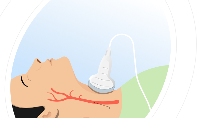
What Is A Carotid Doppler Ultrasound?
Carotid Doppler Ultrasound, also known as a Carotid Duplex Scan, is a diagnostic tool that combines traditional ultrasound and Doppler ultrasound to produce detailed images of the carotid arteries. The traditional ultrasound provides images of the structure of the arteries, while the Doppler ultrasound evaluates the movement of blood within these vessels. This dual capability allows for a comprehensive assessment of both the anatomy and functionality of the carotid arteries. Book a Carotid Doppler Ultrasound in Chandpur for ONLY 1800*
Ultrasound Carotid Doppler Available In
Name
Proc. Time
Rating
Price

Carotid Doppler Ultrasound Purpose
The primary purpose of the Carotid Doppler Ultrasound is to evaluate the health of the carotid arteries, which are crucial for supplying blood to the brain. This test is essential for detecting potential blockages/narrowing (stenosis) that could lead to serious conditions such as strokes. It is particularly valuable for individuals with risk factors like hypertension, diabetes, high cholesterol, or a history of smoking. Additionally, it helps monitor the effectiveness of treatments for carotid artery disease, assess symptoms indicative of impaired blood flow (like sudden numbness, weakness, or visual disturbances), and guide decisions regarding surgical interventions. By providing detailed images and information on blood flow, the Carotid Doppler Ultrasound plays a crucial role in preventive healthcare and the management of cardiovascular diseases. Book a Carotid Doppler Ultrasound in Chandpur for ONLY 1800*
What Does A Carotid Doppler Ultrasound Show?
| Aspect | Details |
|---|---|
| Presence of Blockages | It detects narrowing (stenosis) within the carotid arteries. |
| Plaque Buildup | It identifies accumulation of plaque, indicating atherosclerosis. |
| Blood Flow Speed | It measures the velocity of blood flow through the arteries. |
| Blood Flow Direction | It shows the direction of blood flow, highlighting any abnormal patterns. |
| Blood Clots | It detects presence of blood clots that may pose a stroke risk. |
| Arterial Wall Structure | It provides images of the structure and thickness of the arterial walls. |
| Severity of Stenosis | It assesses the degree of narrowing, indicating the severity of blockages. |
| Treatment Effectiveness | It monitors changes over time to evaluate treatment success. |
| Other Abnormalities | It identifies other potential issues within the carotid arteries. |
Types of Carotid Doppler Ultrasounds
B-mode Ultrasound: This technique produces detailed two-dimensional images of the carotid arteries, providing clear visualisation of the arterial walls and any potential abnormalities such as plaque buildup or structural changes.
Colour Doppler Ultrasound: Using colour mapping, this method shows the direction and speed of blood flow within the carotid arteries. It helps in identifying areas of restricted or turbulent flow, indicating potential blockages or abnormalities.
Spectral Doppler Ultrasound: By measuring the velocity of blood flow, this technique evaluates the severity of stenosis (narrowing) within the carotid arteries. It provides quantitative data on blood flow patterns and helps in assessing the degree of obstruction.
Power Doppler Ultrasound: This method enhances sensitivity to detect blood flow, particularly in small or slow-moving vessels. It is useful for visualising complex blood flow dynamics and identifying subtle changes in flow patterns.
Health Conditions Diagnosed By A Carotid Doppler Ultrasound
Carotid Doppler Ultrasounds are essential for diagnosing and monitoring various health conditions related to the carotid arteries and cardiovascular system:
Carotid Artery Stenosis: This condition involves the narrowing of the carotid arteries due to the buildup of plaque (atherosclerosis). The ultrasound assesses the degree of stenosis, helping determine the risk of stroke. Severe stenosis can significantly reduce blood flow to the brain, increasing the likelihood of ischemic events.
Atherosclerosis: The ultrasound identifies and maps the extent of plaque accumulation on the inner walls of the carotid arteries. Atherosclerosis is a progressive condition that hardens and narrows arteries, leading to reduced blood flow and increased risk of cardiovascular events such as heart attack or stroke.
Blood Clots (Thrombosis): Carotid Doppler Ultrasound detects the presence of blood clots within the carotid arteries. These clots can form due to turbulent blood flow or plaque rupture, posing a severe risk if they dislodge and travel to the brain, causing an ischemic stroke.
Arterial Dissections: This ultrasound technique identifies tears or separations in the carotid artery walls, often caused by trauma or underlying connective tissue disorders. Dissections can obstruct blood flow or lead to the formation of clots, presenting symptoms such as sudden severe headaches, neck pain, or neurological deficits.
Hemodynamic Changes: By assessing blood flow velocity and patterns, Carotid Doppler Ultrasound provides crucial information about vascular health. It helps diagnose conditions such as hypertension, where abnormal blood flow dynamics may indicate increased cardiovascular risk or the presence of vascular disease.
Monitoring Treatment Effectiveness: Carotid Doppler Ultrasound plays a pivotal role in monitoring the effectiveness of treatments aimed at managing carotid artery disease. It evaluates the outcomes of interventions like medication therapy, lifestyle modifications, angioplasty, or stenting, providing insights into disease progression and guiding further therapeutic decisions.
Carotid Doppler Ultrasound plays a crucial role in early detection, ongoing monitoring, and management of these conditions, enabling timely interventions to reduce the risk of stroke and other cardiovascular events. Book a Carotid Doppler Ultrasound in Chandpur for ONLY 1800*
Who Should Get Tested?
Individuals With Risk Factors: Individuals with conditions such as hypertension, diabetes, high cholesterol, obesity, or a history of smoking are recommended to undergo Carotid Doppler Ultrasound. This helps in early detection of carotid artery disease, reducing the risk of stroke.
Patients With Previous Stroke/TIA: Patients who have experienced a stroke or transient ischemic attack (TIA) benefit from Carotid Doppler Ultrasound to assess the extent of artery narrowing. This aids in preventing future strokes by guiding appropriate preventive measures.
Individuals With A Family History Of Stroke Or Carotid Artery Disease: Those with a familial predisposition to stroke or carotid artery disease should consider Carotid Doppler Ultrasound. It helps detect and manage early arterial abnormalities, minimising the risk of cardiovascular complications.
Patients Undergoing Cardiovascular Surgeries: Patients scheduled for heart surgeries or procedures involving the carotid arteries, such as coronary artery bypass grafting (CABG), undergo Carotid Doppler Ultrasound to evaluate artery health and mitigate perioperative stroke risk.
Individuals Presenting Clinical Symptoms: Individuals presenting with symptoms such as unilateral weakness, speech difficulties, or visual disturbances should undergo Carotid Doppler Ultrasound to diagnose carotid artery disease and prevent potential stroke events.
Carotid Doppler Ultrasound plays a pivotal role in the early detection, risk assessment, and management of carotid artery disease, tailored to the specific needs of individuals and patients at risk or experiencing related symptoms. Book a Carotid Doppler Ultrasound in Chandpur for ONLY 1800*
Carotid Doppler Ultrasound Procedure
Preparation: The patient will be asked to lie down on an examination table, usually in a comfortable position. The technologist may ask the patient to remove clothing from the lower limbs or wear a gown for easy access.
Application of Gel: A special gel is applied to the skin over the lower limbs. This gel helps transmit sound waves between the ultrasound probe and the skin, ensuring clear images of the arteries.
Ultrasound Probe Placement: The technologist gently moves an ultrasound probe (transducer) over the gel-covered area of the lower limbs. The probe emits high-frequency sound waves that bounce off tissues and blood cells, creating detailed images of the arteries on a monitor.
Color Doppler Imaging: During the procedure, Color Doppler technology is used to assess blood flow within the arteries. Different colors on the monitor represent the direction and speed of blood flow. This helps identify areas of normal and abnormal blood flow, such as blockages or narrowing.
Doppler Waveform Analysis: The technologist may also perform Doppler waveform analysis. This involves placing the probe at specific points along the arteries to measure the speed and direction of blood flow. Doppler waveforms provide information on the severity of arterial blockages or abnormalities.
Documentation: Images and Doppler waveforms are recorded for review and interpretation by a radiologist or vascular specialist. These findings help in diagnosing vascular conditions and planning appropriate treatments.
Post-Procedure: After the test, the gel is wiped off the skin. There are typically no special instructions or restrictions following the procedure, and patients can resume normal activities.
Results: The results of the Ultrasound Color Doppler Both Lower Limbs Arterial test are usually available shortly after the procedure. A healthcare provider will discuss the findings with the patient and recommend further steps if necessary, such as additional testing or treatment.
This procedure is non-invasive and painless, providing valuable information about the health and function of the arteries in the lower limbs to aid in the diagnosis and management of vascular conditions. Book a Carotid Doppler Ultrasound in Chandpur for ONLY 1800*
What Do The Results Mean?
NORMAL TEST RESULTS
Good Blood Flow: Peak Systolic Velocity (PSV) below 125 cm/s in both common and internal carotid arteries, and End Diastolic Velocity (EDV) below 40 cm/s. These values indicate that blood is flowing smoothly through the arteries without significant obstruction. The PSV represents the highest blood flow speed during heart contraction, while EDV shows the lowest speed when the heart is relaxed. Normal values suggest no narrowing or blockages that could impede blood flow to the brain.
Healthy Vessel Walls: Intima-Media Thickness (IMT) less than 0.9 mm. This measurement assesses the thickness of the inner two layers of the carotid artery wall. A normal IMT suggests that there's no significant thickening of the arterial walls, which is often an early sign of atherosclerosis. It indicates that the arteries are maintaining their structural integrity and flexibility.
Absence Of significant plaque: No plaque or less than 50% stenosis. Plaque is a buildup of fatty deposits, cholesterol, and other substances in the artery walls. The absence of significant plaque or stenosis (narrowing) less than 50% indicates that the arteries are relatively clear and not obstructed. This is crucial for maintaining adequate blood flow to the brain and reducing stroke risk.
No Stenosis: ICA/CCA PSV Ratio less than 2.0. This ratio compares the blood flow velocity in the internal carotid artery (ICA) to that in the common carotid artery (CCA). A ratio below 2.0 confirms that there's no significant narrowing in the internal carotid artery, which is the main blood supply to the brain.
Normal Vessel Elasticity: Resistive Index (RI) between 0.55 and 0.75, and Pulsatility Index (PI) between 0.8 and 1.2. These indices reflect the elasticity and resistance of the blood vessels. Normal values indicate that arteries are appropriately flexible and can accommodate changes in blood flow. The RI measures the resistance to blood flow in the arteries, while the PI assesses the variability of blood velocity in the cardiac cycle.
Balanced Blood Flow Dynamics: The relationship between systolic and diastolic flow, as reflected in the PSV, EDV, RI, and PI values, is normal. This balance indicates that the arteries are managing the pulsatile nature of blood flow, expanding during systole & recoiling during diastole to ensure constant blood supply to the brain.
Symmetrical Findings: Similar normal characteristics in both left and right carotid arteries. This symmetry is important as it suggests that both sides of the brain are receiving comparable blood flow. Significant differences between the two sides could indicate a problem in one of the arteries.
Low stroke Risk: All measurements within normal ranges suggest no increased risk of stroke due to carotid artery issues. Stroke risk is elevated when there's significant stenosis, plaque buildup, or abnormal blood flow in the carotid arteries. Normal findings across all parameters indicate that the carotid arteries are not contributing to an increased stroke risk.
Absence Of Vascular Disease Indicators: Normal values across all measurements indicate no signs of conditions like atherosclerosis, which is characterised by the buildup of plaque in the artery walls. The combination of normal IMT, absence of significant plaque, and normal blood flow velocities suggests healthy arteries without signs of vascular disease.
Age-appropriate Findings: Results are within normal ranges considering the patient age and general health status. It is important to note that some degree of arterial thickening and reduced elasticity is expected with ageing. Normal results in the context of the patient age suggest that any age-related changes are not clinically significant and are within the expected range.
ABNORMAL TEST RESULTS
Impaired Blood Flow: Peak Systolic Velocity (PSV) above 125 cm/s in common or internal carotid arteries, and End Diastolic Velocity (EDV) above 40 cm/s. These elevated values indicate that blood is flowing at higher speeds through the arteries, suggesting narrowing or obstruction. PSV > 230 cm/s often indicates severe stenosis (>70% narrowing). The increased velocity occurs as blood rushes through a constricted area, like water through a narrowed pipe.
Unhealthy Vessel Walls: Intima-Media Thickness (IMT) greater than 0.9 mm. This increased thickness of the inner two layers of the carotid artery wall is often an early sign of atherosclerosis. It suggests that the arteries are losing their structural integrity and flexibility, which can lead to further complications like plaque formation and reduced blood flow.
Presence Of Significant Plaque: Plaque causing more than 50% stenosis. This indicates a substantial buildup of fatty deposits, cholesterol, and other substances in the artery walls. Significant plaque narrows the artery, obstructing blood flow and increasing the risk of complete blockage. The characteristics of the plaque (e.g., ulcerated, heterogeneous) can indicate higher risk of complications.
Stenosis Present: ICA/CCA PSV Ratio is greater than 2.0. This elevated ratio comparing the blood flow velocity in the internal carotid artery to that in the common carotid artery suggests narrowing in the internal carotid artery. Higher ratios correlate with severe stenosis, indicating a greater risk to brain blood supply.
Abnormal Vessel Elasticity: Resistive Index (RI) below 0.55 or above 0.75, and Pulsatility Index (PI) below 0.8 or above 1.2. Abnormal RI might indicate proximal stenosis, hyperperfusion (RI < 0.55), or increased distal resistance (RI > 0.75). Abnormal PI suggests changes in vascular compliance or resistance, with low PI potentially indicating proximal stenosis and high PI suggesting distal resistance.
Imbalanced Blood Flow Dynamics: Abnormal relationship between systolic and diastolic flow, as reflected in the PSV, EDV, RI, and PI values. This imbalance suggests the arteries are not effectively managing the pulsatile nature of blood flow, which leads to irregular blood supply to the brain and more stress on the vessel walls.
Asymmetrical Findings: Significant differences between left and right carotid arteries. This asymmetry is concerning as it suggests that one side of the brain may be receiving less blood flow than the other. It often indicates unilateral disease, which requires further investigation and potentially more urgent intervention.
Increased Stroke Risk: Abnormal measurements, particularly significant stenosis (>50%), elevated velocities, or presence of unstable plaque, indicate an increased risk of stroke. These findings suggest that the carotid arteries are contributing to a higher probability of blood clot formation or decreased blood flow to the brain.
Presence Of Vascular Disease Indicators: Abnormal values across measurements indicate signs of conditions like atherosclerosis. The combination of increased IMT, presence of significant plaque, and abnormal blood flow velocities suggests unhealthy arteries with signs of vascular disease progression.
Age-inappropriate Findings: Results that are outside normal ranges, considering the patient age and general health status. While arterial changes are expected with ageing, abnormal results suggest that the changes are more severe than expected for the patient age, indicating accelerated vascular aging or disease processes.
These abnormal findings collectively paint a picture of compromised carotid artery health, suggesting impaired blood flow to the brain, increased risk of stroke, and the presence of significant vascular disease. They often necessitate further medical evaluation and potential interventions to manage the identified risks and prevent complications. Book a Carotid Doppler Ultrasound in Chandpur for ONLY 1800*
Associated Risks
Discomfort/Pain: Some patients may experience mild discomfort or pressure on the neck during the procedure, especially if the ultrasound transducer is pressed firmly against the skin to obtain clear images.
Allergic Reaction To Gel: The gel used to facilitate ultrasound transmission can rarely cause skin irritation or allergic reactions, particularly in individuals with sensitive skin or known allergies to similar substances.
False Positives Or Negatives: Interpretation of ultrasound images may occasionally lead to false positives (indicating a problem that does not exist) or false negatives (missing an existing problem), depending on factors like operator skill and image quality.
Risk Of Stroke: Although extremely rare, there is a theoretical risk of dislodging plaque during the procedure, which could potentially cause a stroke. This risk is minimal in experienced hands and with proper technique.
Inconclusive Results: Sometimes, ultrasound images may not provide definitive information, requiring additional imaging or tests to confirm or clarify findings.
Exposure To Sound Waves: Although ultrasound uses sound waves instead of radiation, the long-term effects of repeated exposure to ultrasound waves are not fully understood. It is generally considered safe for diagnostic purposes, but precautions are taken to minimise unnecessary exposure.
Tips For Maintaining Cartoid Artery Health
| TIPS | DESCRIPTION |
|---|---|
| Maintain A Healthy Diet | Eat fruits, vegetables, whole grains, and lean proteins. Limit saturated fats and sodium intake. |
| Regular Exercise | Engage in aerobic activities like walking, swimming, or cycling for at least 30 minutes most days. |
| Control Blood Pressure | Monitor blood pressure levels regularly. Manage with diet, exercise, and medications as prescribed. |
| Manage Cholesterol Levels | Avoid high-fat foods, manage weight, and take medications to lower cholesterol levels if necessary. |
| Quit Smoking | Quit smoking to reduce the risk of artery damage and cardiovascular disease. |
| Manage Diabetes | Control blood sugar levels through diet, exercise, and medication management. |
| Maintain A Healthy Weight | Achieve and maintain a healthy weight through balanced eating and regular physical activity. |
| Regular Health Check-ups | Schedule routine visits with healthcare providers to monitor cardiovascular health. |
| Reduce Stress | Practice stress-reducing activities such as meditation, yoga, or deep breathing exercises. |
| Follow Medical Advice | Adhere to prescribed medications, treatment plans, and lifestyle recommendations from healthcare providers. |
Frequently Asked Questions/FAQs
What Is A Carotid Doppler Ultrasound?
Carotid Doppler Ultrasound is an imaging technique used to assess the health of the carotid arteries, which supply blood to the brain. It uses high-frequency sound waves to create images of the arteries and measure blood flow.Why Do People Get A Carotid Doppler Ultrasound?
People undergo Carotid Doppler Ultrasound to evaluate the presence of plaque buildup, detect stenosis, and assess blood flow in the carotid arteries. This identifies potential risks for stroke & other cardiovascular conditions.Where Can I Get A Carotid Doppler Ultrasound Done?
Carotid Doppler Ultrasound is performed at hospitals, diagnostic centres, and specialised clinics with ultrasound capabilities. It is conducted by trained technologists and interpreted by vascular specialists or radiologists.What Does A Carotid Doppler Ultrasound Detect That Other Tests Cannot?
Carotid Doppler Ultrasound specifically assesses the structure and function of the carotid arteries, providing detailed images of plaque buildup, blood flow patterns, and potential narrowing that can lead to stroke.Is A Carotid Doppler Ultrasound Painful Or Uncomfortable?
The procedure is generally painless and non-invasive. You may feel mild pressure from the ultrasound transducer on your neck, but discomfort is minimal.What Should I Do If I Have Concerns About My Carotid Artery Health?
Discuss your concerns with your healthcare provider. They can evaluate your risk factors and determine if further testing, including Carotid Doppler Ultrasound, is appropriate.What Symptoms Indicate The Need For A Carotid Doppler Ultrasound?
Symptoms such as transient ischemic attacks or mini-strokes, sudden weakness/numbness on one body side, difficulty speaking or understanding speech, and vision problems may warrant a carotid doppler ultrasound.How Does A Carotid Doppler Ultrasound Differ From Other Imaging Tests Like CT Scans/MRIs?
Carotid Doppler Ultrasound uses sound waves to create images of blood flow in the carotid arteries, whereas CT scans and MRIs use x-rays and magnetic fields to generate detailed images of structures and tissues in the body.Are There Any Lifestyle Changes That Can Help Improve Carotid Artery Health?
Adopting a healthy lifestyle which includes regular exercise, balanced diets (low in saturated fats and cholesterol), no smoking, and stress management can help improve carotid artery health.Can A Carotid Doppler Ultrasound Detect Blood Clots?
Yes, a carotid doppler ultrasound can detect the presence of blood clots or thrombi in the carotid arteries.How Long Does A Carotid Doppler Ultrasound Take?
The procedure usually takes around 30 to 45 minutes, depending on factors such as the complexity of the imaging needed and the individuals anatomy.Are There Any Age-specific Considerations For Undergoing Carotid Doppler Ultrasound?
While age alone may not dictate the need for a carotid doppler ultrasound, older individuals or those with additional cardiovascular risk factors may be more likely to benefit from screening.How Does Smoking/Tobacco Affect The Test Results?
Smoking and tobacco can contribute to the development and progression of carotid artery disease by promoting plaque buildup and narrowing of the arteries. Quitting smoking improves vascular health and reduces stroke risk.