Overview
Magnetic Resonance Imaging (MRI) is a non-invasive diagnostic tool widely used in modern medicine to provide detailed images of the body’s internal structures. A lower abdominal MRI specifically focuses on the organs, tissues, and structures in the lower abdominal region, offering critical insights for diagnosing various medical conditions. This advanced imaging technique uses powerful magnets and radio waves to produce high-resolution images, helping healthcare professionals in accurate diagnosis and treatment planning.
What Is A Lower Abdominal MRI?
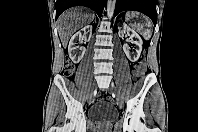
A lower abdominal MRI is an imaging test that captures detailed images of the lower abdominal area, including organs such as the intestines, bladder, prostate (in men), uterus and ovaries (in women), and surrounding tissues. Unlike X-rays and CT scans, which use ionising radiation, MRI uses magnetic fields and radio waves to generate images, making it a safer option for many patients. The images produced by MRI are highly detailed and can show abnormalities that might not be visible with other imaging methods. This procedure is also known by several alternative names, including nuclear magnetic resonance (NMR) - abdomen, magnetic resonance imaging (MRI) – abdomen, and MRI of the abdomen. 5300*

MRI Lower Abdomen Available In
Name
Proc. Time
Rating
Price
What Is Its Purpose?
The primary purpose of a lower abdominal MRI scan is to provide detailed, high-resolution images of the organs, tissues, and structures within the lower abdominal region, facilitating accurate diagnosis and treatment planning. This imaging technique is invaluable for detecting and characterising abnormalities such as tumours, cysts, and inflammatory conditions that might not be visible through other diagnostic methods. Additionally, a lower abdominal MRI helps in evaluating unexplained abdominal pain, assessing the extent of infections, and monitoring chronic diseases like inflammatory bowel disease or cancer. It is also used to guide surgical planning, ensuring precision and reducing surgical risks. By offering a non-invasive and highly detailed examination, a lower abdominal MRI plays a crucial role in improving patient outcomes through early and precise detection of medical issues. 5300*
Why Choose MRIs?
MRI scans are a preferred diagnostic tool for several compelling reasons. They do not use ionising radiation, unlike X-rays and CT scans, which makes them safer for patients. The images produced by MRI are highly detailed, especially for soft tissues, organs, and other structures, enabling the detection of abnormalities that other imaging methods might miss. MRIs are versatile and can be used to examine various parts of the body, including the brain, spine, joints, and abdominal organs. The procedure is non-invasive and generally safe, with minimal side effects. When necessary, contrast agents can enhance image clarity, providing even more diagnostic information. Additionally, MRIs offer superior soft tissue contrast, crucial for diagnosing conditions like tumours, inflammation, and infections. They can also provide functional imaging, such as blood flow and tissue metabolism, offering deeper insights into organ and tissue function. 5300*
Lower Abdominal MRI: With And Without Contrast
An MRI of the lower abdomen can be performed with or without a contrast agent to enhance image clarity.
Abdominal MRI with contrast: uses a gadolinium-based substance administered intravenously to highlight specific tissues and abnormalities. This enhances the detection and characterisation of tumours, vascular issues, and inflammatory or infectious processes, making it ideal for detailed diagnostic and treatment planning.
Abdominal MRI without contrast: on the other hand, relies solely on magnetic resonance technology to provide high-resolution images of the abdominal organs, such as the liver, kidneys, pancreas, intestines, and reproductive organs. It is safe, non-invasive, and suitable for routine screenings and monitoring without the risks associated with contrast agents.
Difference Between Lower Abdominal MRI Scan With & Without Contrast
| Aspect | MRI of Lower Abdomen with Contrast | MRI of Lower Abdomen without Contrast |
|---|---|---|
| Contrast Agent | Uses a gadolinium-based contrast agent to enhance visualisation | No contrast agent used |
| Procedure | Contrast agent administered intravenously to highlight specific tissues | Relies on MRI technology alone |
| Visualisation | Enhances detection of tumours, vascular issues, and inflammatory/infectious processes | Provides detailed images of organs without enhanced contrast |
| Usage | Ideal for detailed diagnostic and treatment planning | Suitable for routine screenings and monitoring |
| Applications | Useful in evaluating and planning treatment for complex conditions | Effective for assessing structural abnormalities and chronic conditions |
| Safety | Generally safe but may have risks like allergic reactions or nephrogenic systemic fibrosis | Safe and non-invasive with no contrast-related risks |
| Patient Suitability | Best for patients needing detailed imaging for diagnosis and treatment | Ideal for those who may be at risk for adverse reactions to contrast agents |
| Imaging Quality | Provides enhanced and detailed anatomical information | Offers high-resolution imaging without contrast enhancement |
MRI Lower Abdomen Available In
| MRI Scans | City | Price | ||
|---|---|---|---|---|
| 1 | MRI Lower Abdomen | - | 5300 |
Lower Abdominal MRI Scan vs. CT Scan
When comparing a lower abdominal MRI scan to a CT scan, several key differences emerge. An MRI scan uses magnetic fields and radio waves, avoiding the ionising radiation exposure that comes with CT scans. This makes MRIs a safer option, especially for patients requiring frequent imaging. MRI provides highly detailed images of soft tissues and organs, which are crucial for detecting abnormalities that CT scans might miss. While MRIs are safe and non-invasive, they do take longer, typically 30-60 minutes, compared to the 5-10 minutes duration of CT scans
CT scans, on the other hand, use ionising radiation, posing a higher risk with repeated exposure but are widely available and cost-effective. They are particularly effective for evaluating bone injuries, lung and chest imaging, and detecting organ injuries. However, contrast agents used in MRIs are gadolinium-based and less likely to cause allergic reactions, whereas CT scans use iodine-based contrast, which can trigger allergies. Despite the duration and potential discomfort for claustrophobic patients, the detailed imaging makes MRIs better for evaluating soft tissues, tumours, inflammation, and vascular issues.5300*
Diseases Diagnosed By Lower Abdominal MRI
A lower abdominal MRI is a powerful diagnostic tool that can detect and evaluate a wide range of diseases and conditions affecting the lower abdominal region. The detailed images produced by this scan allow healthcare providers to diagnose and monitor various medical issues with high accuracy.
Tumours and Cancers: MRI scans are highly effective in detecting and staging cancers in the lower abdomen, including colorectal, bladder, ovarian, and prostate cancers. These scans identify masses, differentiate between benign and malignant tumours, and monitor the progression to treatment of these cancers.
Inflammatory and Infectious Conditions: Lower abdominal MRI can accurately diagnose conditions like appendicitis, which requires prompt surgical intervention, and inflammatory bowel diseases such as Crohn’s disease and ulcerative colitis. It also evaluates pelvic inflammatory disease (PID) and diverticulitis by detecting inflammation and infection in the intestines and reproductive organs.
Structural Abnormalities: MRI is useful in identifying structural abnormalities such as hernias (inguinal or femoral), uterine fibroids, and other uterine anomalies. It also detects endometriosis, which involves the presence of endometrial tissue outside the uterus, and hydronephrosis, which is the swelling of a kidney due to urine buildup caused by urinary tract obstruction.
Vascular Conditions: This imaging technique is essential for detecting vascular conditions like abdominal aortic aneurysms, which are abnormal bulges in blood vessels, and deep vein thrombosis (DVT), which involves blood clots in the veins of the lower abdomen and pelvis. It also evaluates mesenteric ischemia by assessing blood flow to the intestines and detecting restricted blood supply.
Urinary Tract Issues: Lower abdominal MRI can detect kidney stones in the kidneys, ureters, or bladder. It is also effective in evaluating interstitial cystitis, a chronic inflammation of the bladder wall, and assessing structural issues contributing to urinary incontinence.
A lower abdomen MRI is particularly useful for diagnosing complex and serious conditions, ensuring patients receive precise diagnoses and appropriate treatments. 5300*
Who Should Get Tested?
{ "Individuals Experiencing Persistent Lower Abdominal Pain": { "description": "Individuals with persistent lower abdominal pain that does not improve with conservative treatments may benefit from a lower abdominal MRI to identify potential causes such as gastrointestinal disorders, reproductive organ abnormalities, or urinary tract issues." }, "Patients Presenting with Gastrointestinal Symptoms": { "description": "Patients with gastrointestinal symptoms such as bloating, diarrhea, constipation, or unexplained weight loss may require a lower abdominal MRI to evaluate the intestines, stomach, and other digestive organs for conditions such as inflammatory bowel disease, tumours, or bowel obstructions." }, "Individuals with Suspected Kidney or Bladder Conditions": { "description": "Individuals with symptoms suggestive of kidney or bladder issues, such as hematuria (blood in the urine), frequent urination, or flank pain, may undergo a lower abdominal MRI to assess the kidneys, ureters, bladder, and surrounding structures for abnormalities such as stones, tumours, or infections." }, "Patients with Reproductive Health Concerns": { "description": "Patients with reproductive health concerns such as pelvic pain, abnormal bleeding, or infertility may benefit from a lower abdominal MRI to evaluate the reproductive organs (e.g., uterus, ovaries, fallopian tubes) and detect conditions such as fibroids, ovarian cysts, or endometriosis." }, "Individuals Undergoing Cancer Screening or Monitoring": { "description": "Individuals with a history of cancer or at risk of developing abdominal tumours may require periodic screening or monitoring with a lower abdominal MRI to detect and evaluate tumours in organs such as the liver, pancreas, or adrenal glands." }, "Patients with Chronic Conditions": { "description": "Patients with chronic conditions affecting the lower abdomen, such as Crohn's disease, ulcerative colitis, or polycystic kidney disease, may undergo regular lower abdominal MRIs to monitor disease progression, assess treatment response, and detect complications." }, "Individuals Undergoing Preoperative Planning or Postoperative Assessment": { "description": "Individuals scheduled for abdominal surgery, such as organ transplantation or tumour resection, may undergo a lower abdominal MRI for preoperative planning to evaluate the extent of pathology and assist in surgical decision-making. Similarly, postoperative assessment with a lower abdominal MRI may be necessary to evaluate surgical outcomes, detect any postoperative complications, and guide further management." }}
0:
1:
2:
3:
4:
5:
6:
7:
8:
9:
10:
11:
12:
13:
14:
15:
16:
17:
18:
19:
20:
21:
22:
23:
24:
25:
26:
27:
28:
29:
30:
31:
32:
33:
34:
35:
36:
37:
38:
39:
40:
41:
42:
43:
44:
45:
46:
47:
48:
49:
50:
51:
52:
53:
54:
55:
56:
57:
58:
59:
60:
61:
62:
63:
64:
65:
66:
67:
68:
69:
70:
71:
72:
73:
74:
75:
76:
77:
78:
79:
80:
81:
82:
83:
84:
85:
86:
87:
88:
89:
90:
91:
92:
93:
94:
95:
96:
97:
98:
99:
100:
101:
102:
103:
104:
105:
106:
107:
108:
109:
110:
111:
112:
113:
114:
115:
116:
117:
118:
119:
120:
121:
122:
123:
124:
125:
126:
127:
128:
129:
130:
131:
132:
133:
134:
135:
136:
137:
138:
139:
140:
141:
142:
143:
144:
145:
146:
147:
148:
149:
150:
151:
152:
153:
154:
155:
156:
157:
158:
159:
160:
161:
162:
163:
164:
165:
166:
167:
168:
169:
170:
171:
172:
173:
174:
175:
176:
177:
178:
179:
180:
181:
182:
183:
184:
185:
186:
187:
188:
189:
190:
191:
192:
193:
194:
195:
196:
197:
198:
199:
200:
201:
202:
203:
204:
205:
206:
207:
208:
209:
210:
211:
212:
213:
214:
215:
216:
217:
218:
219:
220:
221:
222:
223:
224:
225:
226:
227:
228:
229:
230:
231:
232:
233:
234:
235:
236:
237:
238:
239:
240:
241:
242:
243:
244:
245:
246:
247:
248:
249:
250:
251:
252:
253:
254:
255:
256:
257:
258:
259:
260:
261:
Ultimately, the decision to undergo a lower abdominal MRI should be made in consultation with a healthcare provider, who can assess the individual's medical history, symptoms, and diagnostic needs to determine the most appropriate imaging approach. 5300*
How A Lower Abdominal MRI Works
A lower abdominal MRI scan is a sophisticated imaging technique that provides detailed pictures of the structures within the lower abdomen. It works by using a combination of powerful magnets, radio waves, and computer technology to create high-resolution
Here’s a detailed look at how this scan works:
Magnetic Field Creation: The MRI machine generates a strong magnetic field that temporarily realigns hydrogen atoms in the body’s tissues. The human body is composed largely of water, which contains hydrogen atoms. These atoms are key to the MRI process.
Radio Wave Pulses: Once the hydrogen atoms are aligned, the machine sends out radiofrequency (RF) pulses. These pulses knock the hydrogen atoms out of alignment.
Resonance and Signal Detection: When the RF pulses are turned off, the hydrogen atoms return to their original alignment. As they do, they emit energy signals. Different tissues in the abdomen (such as organs, muscles, and fat) return to their original states at different rates, producing distinct signals.
Signal Capture: The MRI machine detects these energy signals using coils placed around the abdomen. The coils capture the signals emitted by the hydrogen atoms as they return to their original alignment.
Image Formation: The signals are then processed by a computer to create detailed cross-sectional images of the abdominal structures. These images can be viewed in various planes (sagittal, coronal, and axial) and provide comprehensive views of the internal organs, blood vessels, and other tissues.
Contrast Enhancement (if used): In some cases, a contrast agent (typically gadolinium-based) is injected into the patient’s bloodstream before the scan. The contrast agent highlights specific areas, such as blood vessels or abnormal tissues, making them more visible on the MRI images. The contrast works by altering the local magnetic environment, thereby enhancing the signal difference between various tissues.
Image Review: The generated images are reviewed by a radiologist, a doctor specialised in interpreting medical images. The radiologist looks for abnormalities or changes in the structures of the lower abdomen, aiding in the diagnosis of various conditions.
The lower abdominal MRI scan provides highly detailed images that can reveal a wide range of conditions, from structural abnormalities to tumours and inflammatory diseases, making it an invaluable tool in modern medical diagnostics. For any abdominal health concerns, ensuring timely testing can make a significant difference. Visit www.flebo.in and get tested in time. 5300*
Lower Abdominal MRI Procedure
A lower abdominal MRI scan is a comprehensive imaging technique used to examine the structures within the lower abdomen. Here’s an overview of the procedure:
Preparation: Before the scan, patients remove all metal objects, change into a hospital gown, and may need to provide information about any metal implants or medical conditions.
Positioning: The patient lies flat on their back on the MRI table, with cushions and straps used for comfort and to minimise movement. Proper positioning is crucial for obtaining clear, accurate images.
Contrast Injection: If required, a gadolinium-based contrast dye is injected into a vein to enhance image clarity. The patient is monitored for any allergic reactions to the contrast agent.
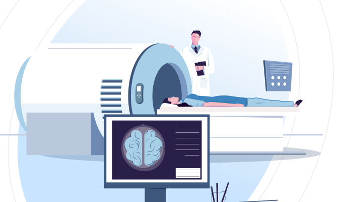
Entering The MRI Machine: The table slides into the MRI machine. The machine produces loud noises during the scan, so patients are provided with earplugs or headphones to minimise discomfort from the noise.
Scanning Process: The patient remains still as the MRI machine captures detailed images using magnets and radio waves. The procedure takes 30 to 60 minutes, depending on whether contrast is used.
Results: A radiologist reviews the images and sends a report to the referring doctor. The doctor will then discuss the findings and any necessary follow-up steps with the patient.
This detailed procedure provides critical insights into the lower abdominal region, aiding in the diagnosis and management of various medical conditions. 5300*
NORMAL TEST RESULTS
A normal result from a lower abdominal MRI scan indicates that the internal structures and organs within the lower abdominal region appear healthy and there are no detectable abnormalities.
Here’s what a normal result generally signifies:
Absence of Tumours or Masses: No abnormal growths, tumours, cysts, or masses are present in the lower abdominal organs such as the liver, kidneys, bladder, intestines, and reproductive organs.
Normal Organ Size and Shape: Organs within the lower abdomen, including liver, kidneys, bladder, pancreas, and reproductive organs, are of normal size and shape, with no enlargement, shrinkage, or displacement.
Healthy Tissue Appearance: The tissues of the lower abdominal organs show a normal appearance with no signs of abnormal signal intensity that would suggest inflammation, infection, or other pathological changes.
Clear Blood Vessels: The blood vessels in the lower abdomen, including arteries and veins, appear normal with no signs of aneurysms, blockages, or abnormal blood flow patterns.
Absence of Fluid Collections: There are no abnormal fluid collections such as abscesses, cysts, or free fluid within the abdominal cavity, which might indicate an underlying infection or other medical condition.
No Structural Abnormalities: The MRI images do not show any structural abnormalities or congenital anomalies in the lower abdominal organs and surrounding structures.
Normal Lymph Nodes: The lymph nodes in the lower abdominal region are of normal size and appearance, with no signs of enlargement or other abnormalities that suggest infection, inflammation, or malignancy.
A normal result from a lower abdominal MRI scan indicates that the internal structures and organs within the lower abdominal region appear healthy and there are no detectable abnormalities. 5300*
ABNORMAL TEST RESULTS
An abnormal result from a lower abdomen MRI indicates that the imaging has revealed some irregularities orconcerns within the scanned area. Here’s a breakdown of what different types of abnormal findings might mean:
Tumours Or Masses: The presence of abnormal growths, which could be benign (non-cancerous) or malignant (cancerous). These masses might be found in organs such as the liver, kidneys, bladder, intestines, or reproductive organs.
Organ Abnormalities: Changes in the size, shape, or position of organs. This could include enlarged organs (hepatomegaly for the liver, splenomegaly for the spleen), shrunken organs, or organs that are not in their usual location (organ displacement).
Abnormal Tissue Signals: Areas that show unusual signal intensity might indicate inflammation, infection, fibrosis, or other pathological changes. For example, inflamed bowel walls could suggest conditions like Crohn’s disease or ulcerative colitis.
Fluid Collections: Detection of abnormal fluid collections such as abscesses, cysts, or ascites (fluid in the abdominal cavity). These could indicate infections, inflammatory conditions, or other medical issues.
Blood Vessel Abnormalities: Findings such as aneurysms (abnormal bulges in blood vessels), blockages, or abnormal blood flow patterns. These could be related to conditions like vascular diseases or blood clots.
Lymph Node Changes: Enlarged or irregular lymph nodes can indicate infections, inflammation, or malignancies. Enlarged lymph nodes may be a sign of lymphoma or metastasis from another cancer.
Structural Abnormalities: Congenital anomalies (present from birth) or acquired structural issues, such as hernias or adhesions (scar tissue bands), which might affect organ function or cause pain.
Signs Of Trauma: Evidence of past injuries such as fractures, organ lacerations, or hematomas (localised blood collection), which might not have fully healed or might be causing ongoing issues.
An abnormal result from a lower abdomen MRI indicates that the imaging has revealed some irregularities orconcerns within the scanned area. Here’s a breakdown of what different types of abnormal findings might mean: 5300*
Associated Risks
A lower abdominal MRI is generally considered safe and non-invasive, but like any medical procedure, it carries some associated risks and considerations. Here are the primary risks and potential issues:
Discomfort and Claustrophobia: The enclosed space can cause discomfort or claustrophobia. Sedation or alternative imaging methods may be needed.
Contrast Agent Reactions: Small risk of allergic reactions to gadolinium-based contrast agents, ranging from mild (itching) to severe. Patients should inform their provider of any known allergies.
NSF Risk: Patients with severe kidney disease may risk nephrogenic systemic fibrosis (NSF) if gadolinium is used. Screening and careful consideration are necessary to mitigate this risk.
Magnetic Field Effects: Metal implants or devices (e.g., pacemakers) can be affected by the magnetic field. Inform the provider of any implants.
Pregnancy Considerations: MRI is generally safe during pregnancy, but gadolinium contrast is usually avoided unless necessary due to potential risks to the fetus. Non-contrast MRI is preferred.
Noise: Loud noises can be uncomfortable. Earplugs or headphones are provided.
Motion Artefacts: Movement during the scan can blur images, potentially requiring a repeat scan. Patients are advised to remain as still as possible.
Long Duration: MRI scans can be lengthy, which may be uncomfortable for some patients who need to lie still for an extended period.
Inadvertent Heating: In rare cases, the MRI's radiofrequency energy can cause slight body heating. This is usually minimal and not harmful, but it can be uncomfortable.
Precautions To Avoid Abdomen-Related Issues
Healthy Diet: Consuming a balanced diet rich in fruits, vegetables, whole grains, lean proteins, and healthy fats is vital for abdominal health. Avoiding processed foods and sugary drinks can prevent many abdominal issues, while staying hydrated supports digestion.
Regular Exercise: Regular physical activity, including aerobic and strength training exercises, helps maintain a healthy weight and supports digestive health, reducing the risk of abdominal diseases.
Moderate Alcohol Consumption: Limiting alcohol intake reduces the risk of liver disease and other abdominal problems, making moderation crucial for abdominal health.
Avoid Smoking: Smoking increases the risk of cancers, including those affecting the abdomen. Quitting or avoiding smoking significantly reduces this risk.
Regular Medical Check-ups: Regular check-ups enable early detection and monitoring of health issues. Following your doctor’s screening recommendations can catch problems early, ensuring better outcomes.
Avoid Overuse of Medications: Using medications like NSAIDs and antibiotics only as prescribed prevents harm to the stomach and intestines, reducing the risk of abdominal problems.
Maintain Good Posture: Good posture reduces the risk of abdominal discomfort and digestive issues by supporting abdominal muscles and organs, contributing to better overall health.
An abnormal result from a lower abdomen MRI indicates that the imaging has revealed some irregularities orconcerns within the scanned area. Here’s a breakdown of what different types of abnormal findings might mean: 5300*


