
What Is An ECG/EKG?
An ECG or Electrocardiogram, is a diagnostic test that measures and records the electrical activity of the heart over a period of time. By placing small electrodes on the skin, typically on the chest, arms, and legs, the test detects the electrical signals generated by the heart contractions and relaxations. These signals are then displayed graphically as waves on an electrocardiogram, providing healthcare professionals with valuable information about the hearts rhythm and function. ECGs are widely used to diagnose heart conditions, assess cardiac health, and monitor the effects of treatments or medications on the heart. BOOK AN ECG SCAN FOR ₹200*
ECG Available In
Name
Proc. Time
Rating
Price

What Is An ECG Scan Used For?
Monitoring Of Heart Health: ECGs are used to monitor heart health over time, tracking changes in electrical patterns and detecting any new abnormalities that may develop. They help healthcare providers monitor patients with known heart conditions or individuals at risk of cardiovascular disease.
Evaluation Of Symptoms: ECGs assist in evaluating symptoms such as chest pain, shortness of breath, palpitations, dizziness, or fainting episodes. By examining the ECG tracing, healthcare providers can identify the underlying cause of symptoms and determine appropriate treatment strategies.
Screening For Heart Disease: ECGs can be used for screening purposes in asymptomatic individuals with risk factors for cardiovascular disease, such as hypertension, diabetes, smoking, or a family history of heart disease. Screening ECGs help detect early signs of heart abnormalities and allow for timely intervention.
Assessment Of Treatment Response: ECGs are used to monitor the effectiveness of treatments, medications, or interventions aimed at managing cardiac conditions. Changes in ECG patterns over time can indicate whether treatment strategies are successful or if adjustments are needed to optimize patient care.
Guidance For Medical Procedures: ECGs guide medical procedures such as cardiac catheterization, pacemaker implantation, cardioversion, or electrophysiological studies by providing valuable information about the heart's electrical activity and guiding the placement of electrodes or catheters.
Types Of ECG Tests
Resting ECG: This is the standard ECG test wherein electrodes are placed on the chest, arms, and legs, and the patient lies still for a short period while the ECG machine records the heart's electrical activity.
Exercise Stress Test: Known as a treadmill test or exercise ECG, this monitors the heart's response to physical activity. The patient walks on a treadmill or pedals a stationary bike while connected to an ECG machine. The test evaluates heart function during exercise, detecting abnormalities that may not be present at rest.
Holter Monitor: A Holter monitor is a portable device worn by the patient for 24 to 48 hours or longer to continuously record the heart's electrical activity. It provides a more extended period of monitoring, allowing for the detection of intermittent arrhythmias or other abnormalities that may occur during daily activities.
Event Monitor: Similar to a Holter monitor, an event monitor is a portable device worn by the patient for an extended period, ranging from several days to weeks. It is activated by the patient when they experience symptoms such as palpitations or dizziness, recording ECG tracings during symptomatic episodes.
Ambulatory ECG Monitoring: This involves wearing a portable ECG device for an extended period, typically 24 hours or longer, to monitor the heart's electrical activity during normal daily activities and sleep. Ambulatory ECG monitoring helps diagnose arrhythmias, evaluate symptoms, and assess the effectiveness of treatment.
Signal-Averaged ECG: This specialized ECG test is used to detect subtle abnormalities in the heart's electrical signals that may indicate an increased risk of arrhythmias or sudden cardiac death. It involves recording multiple ECG tracings and averaging them to enhance the detection of low-amplitude signals.
Cardiac Event Recorder: Similar to an event monitor, a cardiac event recorder is a portable device that the patient wears for an extended period to record ECG tracings during symptomatic episodes. However, it has longer recording capabilities, allowing for continuous monitoring and storage of data for medical review later.
Who Should Get Tested?
Here are some groups of people who should consider getting tested:
Individuals With Cardiovascular Risk:
Hypertension (high blood pressure)
Hyperlipidemia (high cholesterol)
Diabetes mellitus
Obesity
Smoking
Family history of heart disease
Patients With Heart Disease Symptoms:
Chest pain or discomfort
Shortness of breath
Palpitations (rapid or irregular heartbeats)
Dizziness or lightheadedness
Fainting or syncope
Fatigue or weakness
Individuals With Known Heart Conditions:
Previous myocardial infarction (heart attack)
Coronary artery disease
Heart failure
Arrhythmias (e.g., atrial fibrillation, ventricular tachycardia)
Structural heart abnormalities (e.g., hypertrophic cardiomyopathy, valvular heart disease)
Patients Undergoing Surgery:
Patients undergoing surgery, especially procedures involving anaesthesia or significant stress on the cardiovascular system, may require preoperative ECG testing to assess their cardiac health and identify any potential risks or contraindications to surgery.
Asymptomatic Individuals:
Age (especially over 40-50 years)
Family history of premature heart disease
Sedentary lifestyle
Poor dietary habits
High stress levels
Occupational exposure to cardiovascular risk factors
Individuals Getting Tested:
ECG testing may be included as part of routine health check-ups or preventive care, especially for individuals with multiple risk factors or a history of cardiovascular disease.
Athletes Or Sports Participants:
Athletes or individuals participating in competitive sports may undergo pre-participation screening, which may include ECG testing, to assess their cardiac health and identify any underlying heart conditions that could increase the risk of sudden cardiac events during physical activity. Book an ECG Test in <city> for ONLY <price>
How To Read An ECG Scan
Understand The Paper: ECG tracings are recorded on graph paper with each large square representing 0.2 seconds horizontally and 0.5 millivolts (mV) vertically.
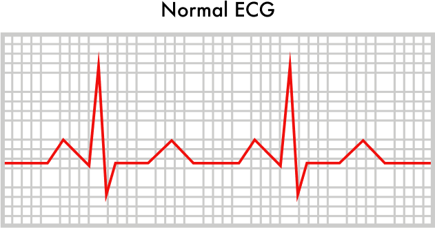
Identify The Basic Waves: ECG waves include the P wave (atrial depolarization), QRS complex (ventricular depolarization), and T wave (ventricular repolarization), each representing a phase of the heart's electrical cycle.
Assess The Heart Rate: Calculate the heart rate by counting the number of QRS complexes in a six-second strip and multiplying by 10 or using the 300, 150, 100, 75 method to estimate heart rate based on R wave intervals.
Evaluate The Rhythm: Determine if the rhythm is regular or irregular by assessing the regularity of RR intervals, indicating potential arrhythmias like atrial fibrillation or flutter.
Check For Intervals And Segments: Examine the PR interval (0.12 to 0.20 seconds), QRS duration (less than 0.12 seconds), and QT interval (adjusted for heart rate), assessing for abnormalities like prolonged QT or PR intervals.
Look For Abnormalities: Identify deviations such as ST segment elevation or depression, T wave abnormalities, and arrhythmias/conduction abnormalities, considering potential myocardial infarction, ischemia, arrhythmias, etc.
Consider Clinical Context: Interpret ECG findings in conjunction with the patient's clinical presentation, medical history, symptoms, and other diagnostic tests for a comprehensive assessment of cardiac health.
How Is This Test Done?
An ECG is a non-invasive procedure that is relatively simple and painless. Here how the test is typically done:
Preparation: The patient is asked to remove any clothing from the waist up and to lie down on an examination table or bed. If necessary, the patient may need to remove jewellery, watches, or other metal objects that could interfere with the ECG recording.
Placement Of Electrodes: Small, adhesive electrodes are placed on the patient's chest, arms, and legs. The number of electrodes used may vary, but the standard ECG involves placing 10 electrodes in specific locations. The skin is cleaned with alcohol pads before electrode placement to ensure optimal signal transmission.
Connection To ECG Machine: The electrodes are connected to the ECG machine via wires or cables. The machine detects and records the electrical signals produced by the heart and displays them as a series of waves on graph paper or a computer screen.
Recording: The ECG technician or healthcare provider initiates the recording process, and the patient is asked to lie still and relax while the ECG is being performed. It typically takes a few minutes to complete the recording, during which time the patient should avoid talking or moving excessively to minimize artifacts.
Analysis: Once the recording is complete, the ECG tracing is analyzed by the healthcare provider or a trained technician. They examine the various waves, intervals, and segments of the ECG tracing to assess the heart's electrical activity, identify any abnormalities, and make a diagnosis if necessary.
Interpretation: The ECG findings are interpreted in the context of the patient's symptoms, medical history, and other diagnostic tests. The results help determine the presence of heart conditions such as arrhythmias, ischemia, myocardial infarction, conduction abnormalities, or structural heart disease.
Documentation: The ECG recording and interpretation are documented in the patient's medical records for future reference. The results may be shared with the patient and healthcare providers involved in their care.
How To Prepare For The Test?
Preparing for an ECG test involves minimal effort however, here are some general guidelines to follow:-
Clothing: Wear a shirt that can be easily lifted or removed as you may need to expose your chest.
Avoid Lotions: Refrain from applying lotions or oils to your chest as they can interfere with electrode adhesion.
Medications: Take your prescribed medications as usual unless advised otherwise by your healthcare provider.
Inform Provider: Share any medical conditions or allergies with your healthcare provider before the procedure.
Relax: Stay calm during the test to ensure accurate results. Inform the provider if you feel anxious or stressed.
Follow Instructions: Adhere to any specific instructions provided by the testing facility for optimal preparation.
What Do You Mean By A Normal ECG Result?
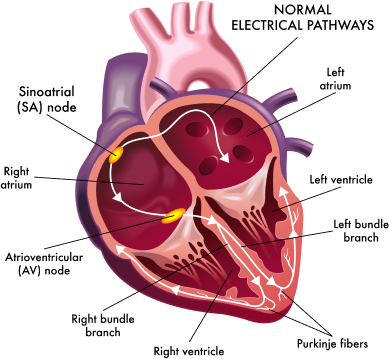
Normal Sinus Rhythm: The rhythm is regular, with consistent intervals between successive QRS complexes. Each QRS complex is preceded by a P wave, and each P wave is followed by a QRS complex. This pattern indicates normal sinus rhythm, originating from the sinoatrial (SA) node.
Heart Rate: The heart rate falls within the normal range, typically between 60 to 100 beats per minute (bpm) at rest for adults. In pediatric populations, the normal heart rate may vary based on age.
P Waves: P waves are present and uniform in appearance, indicating atrial depolarization (contraction). They should be upright in most leads and consistent in morphology across leads.
PR Interval: The PR interval measures the time from the beginning of the P wave to the beginning of the QRS complex. It falls within the normal range of 120 to 200 ms, representing normal atrioventricular conduction.
QRS Complex: QRS complexes are narrow (typically < 120 ms in duration) and consistent in morphology. They represent ventricular depolarization (contraction) and should be upright in most leads.
ST Segment and T Wave: The ST segment is isoelectric (neither elevated nor depressed) and follows the QRS complex. T waves are typically upright and symmetric, representing ventricular repolarization (relaxation).
QT Interval: The QT interval represents ventricular depolarization and repolarization. It varies based on heart rate and falls within the normal range when corrected for heart rate using formulas such as the QTc interval.
Additional Parameters: Other ECG parameters, such as axis deviation, intervals, and waveforms, should also fall within normal ranges based on age, sex, and patient characteristics.
| Normal ranges of ECG parameters | |
|---|---|
| ECG parameter | Normal Range |
| Heart rate | 60-100 beats per minute |
| PR interval | 120-200 milliseconds |
| QRS complex | <120 milliseconds |
| ECG = electrocardiogram. | |
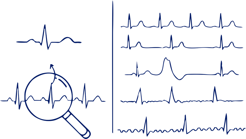
What Do You Mean By Abnormal ECG Result?
Arrhythmias: Irregularities in the heart rhythm, such as atrial fibrillation, atrial flutter, ventricular tachycardia, and bradycardia manifest as irregular intervals between successive QRS complexes or absence of P waves.
ST-Segment Changes: Deviations in the ST segment from the baseline (isoelectric line), including ST-segment elevation or depression. These changes may indicate myocardial ischemia, injury, or infarction.
T-Wave Abnormalities: Inverted, flattened, or peaked T waves may suggest myocardial ischemia, electrolyte imbalances, myocardial injury, or other cardiac abnormalities.
QT Interval Prolongation: Prolongation of the QT interval, corrected for heart rate (QTc), may predispose individuals to ventricular arrhythmias like torsades de pointes. Shortening of the QT interval may also occur.
Bundle Branch Blocks: Delayed/blocked conduction via the bundle branches of the heart, resulting in widened QRS complexes (> 120 ms). This includes left bundle branch block (LBBB) or right bundle branch block (RBBB).
Axis Deviation: Deviation from the normal electrical axis of the heart, which may indicate structural heart disease or conduction abnormalities.
Pathological Q Waves: Abnormal Q waves with significant depth (> 25% of the subsequent R wave) and duration (> 0.04 seconds) may suggest myocardial infarction or scar tissue from previous myocardial injury.
Conduction Abnormalities: Abnormalities in the PR interval, such as prolonged PR interval (> 200 ms) or absence of PR interval (e.g., in atrial fibrillation).
Hypertrophy Patterns: Increased voltage or changes in the morphology of the QRS complexes may indicate ventricular hypertrophy or atrial enlargement.
Other Abnormal Findings: Additional abnormalities such as artifact, lead misplacement, poor electrode contact, or technical errors may also contribute to an abnormal ECG result.
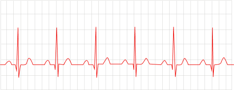
Normal Heartbeat
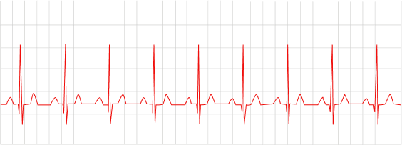
Fast Heartbeat
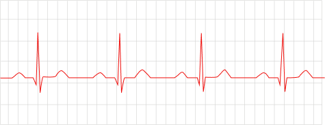
Slow Heartbeat
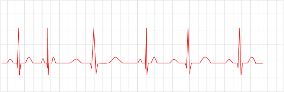
Irregular Heartbeat
| Analysis content | Normal | Abnormal |
|---|---|---|
| Heart rate | 60-100 per minute | < 60 per minute, > 100 per minute |
| Heart rhythm | regular | Irregular |
| P wave | sinus P wave | Non-sinus P wave |
| PR interval Period | 0.12-0.20 seconds | < 0.12 seconds, > 0.20 seconds |
| QRS wave | Normal QRS wave | Abnormal voltage, Abnormal electric axis, QRS duration augmentation, Pathological Q wave |
| ST segment | Normal ST segment | Elevation & depression of ST segment |
| T wave | Normal T wave | Tip, flat, or inverted T wave |
| Other issues | - | U wave, Abnormal electrolyte-related ECG, Drug-related ECG |
Risks Associated With An ECG Scan
Skin Irritation: Adhesive electrodes used to attach to the skin may cause mild irritation or redness, particularly in individuals with sensitive skin. This discomfort is temporary and resolves after removing the electrodes.
Allergic Reaction: Rarely, individuals may experience an allergic reaction to the adhesive material used in the electrodes. Symptoms may include itching, rash, or hives at the electrode sites. If you have a known allergy to adhesives or metals, inform your healthcare provider before the test.
Electrode Discomfort: Some individuals may experience minor discomfort or pressure at the electrode sites during the test, particularly if the electrodes are applied too tightly or if the skin is sensitive.
False Positive or Negative Results: While ECGs are valuable diagnostic tools, they are not infallible. In some cases, ECGs produce false positive or negative results, leading to incorrect interpretations of cardiac health.
Rare Complications: In extremely rare cases, individuals may experience complications such as fainting (vasovagal syncope) or cardiac arrhythmias during the test. These complications are exceptionally uncommon and typically occur in individuals with underlying cardiac conditions.