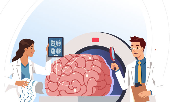
What Is A Brain MRI?
Brain MRI, short for Magnetic Resonance Imaging of the Brain, is a sophisticated medical imaging technique that utilises powerful magnets and radio waves to generate detailed images of the brain’s structure and function. Unlike other imaging methods such as X-rays or CT scans, a brain MRI provides high-resolution, three-dimensional images without using ionising radiation, making it safe for repeated use, especially for soft tissue visualisation. Currently, MRIs are the most sensitive imaging scan of your head (particularly, your brain), as compared to other imaging techniques. Book MRI Brain in Hooghly for2750*
Purpose Of A Brain MRI
The primary purpose of a brain MRI is to provide detailed diagnostic images of the brain’s structure and function. It helps healthcare professionals identify and assess neurological conditions such as tumours, strokes, infections, multiple sclerosis, and neurodegenerative diseases. Additionally, it guides treatment decisions and monitors disease progression. Beyond its clinical utility, a brain MRI also fuels scientific research by uncovering the brain’s anatomy, connectivity, and pathology, contributing to advancements in neuroscience and patient care.
MRI Brain Available In
Name
Proc. Time
Rating
Price

What Does A Brain MRI Show?
A brain MRI provides detailed images of the brain internal structures, allowing healthcare professionals to visualise and evaluate various anatomical and pathological features. Here what a brain MRI can show:
Brain Anatomy: MRI produces high-resolution images that depict the brain anatomy in detail, including the cerebral cortex, white matter, basal ganglia, brainstem, and cerebellum. It allows visualisation of different brain structures and their spatial relationships.
Lesions and Abnormalities: Brain MRI can detect a wide range of lesions and abnormalities, including tumours, cysts, abscesses, hematomas, infarcts (strokes), and demyelinating plaques. These abnormalities appear as areas of altered signal intensity on MRI images.
Tumour Characteristics: MRI helps characterise brain tumours by assessing their location, size, shape, and internal features. It distinguishes between different tumour types (e.g., gliomas, meningiomas) based on their appearance on various MRI sequences (e.g., T1-weighted, T2-weighted, contrast-enhanced images).
Vascular Structures: MRI can visualise blood vessels in the brain using techniques such as magnetic resonance angiography (MRA) or magnetic resonance venography (MRV). It helps assess vascular abnormalities such as aneurysms, arteriovenous malformations (AVMs), stenosis, or occlusions.
Ischemic Stroke: MRI is highly sensitive for detecting ischemic stroke (caused by a blockage in a blood vessel). It shows areas of restricted diffusion on diffusion-weighted imaging (DWI), indicating acute infarction, as well as corresponding changes on other sequences.
Hemorrhagic Stroke: MRI is essential for diagnosing hemorrhagic stroke (caused by bleeding in the brain). It visualises acute haemorrhage as areas of hyperintensity on gradient-echo sequences (e.g., T2*-weighted or susceptibility-weighted imaging).
Multiple Sclerosis (MS) Lesions: Brain MRI is a key tool for diagnosing and monitoring multiple sclerosis (MS). It shows characteristic MS lesions (plaques) in the brain and spinal cord, which appear as hyperintense areas on T2-weighted and fluid-attenuated inversion recovery (FLAIR) images.
Brain Atrophy: MRI can assess for brain atrophy, which is characterised by a loss of brain tissue volume. It may occur as a result of ageing, neurodegenerative diseases like Alzheimer and frontotemporal dementia, or other pathological processes.
Infections and Inflammatory Conditions: Brain MRI can detect features of brain infections (e.g., encephalitis, meningitis) and inflammatory conditions (e.g., autoimmune encephalitis, demyelinating diseases). It shows signs of inflammation, oedema, enhancement, or abscess formation.
Structural Anomalies: MRI reveals structural anomalies of the brain, including congenital malformations, developmental abnormalities, or abnormalities of the pituitary gland or pineal gland.
Overall, brain MRI provides valuable diagnostic information about the brain structure, function, and pathology, helping guide clinical management and treatment decisions for various neurological conditions. Book MRI Brain in Hooghly for2750*
How Does A Brain MRI Work?
A Brain MRI works by utilising a powerful magnetic field, radio waves, and a sophisticated computer system to generate detailed images of the brain’s internal structures. Here’s how it works:-
Magnetic Field Alignment: When a patient enters the MRI machine, they are exposed to a strong magnetic field generated by the MRI scanner. This magnetic field causes the hydrogen atoms in the water molecules within the body’s tissues, including the brain, to align in a specific direction.
Radiofrequency Pulses: Radiofrequency coils within the MRI scanner emit short bursts of radio waves, which are directed at the aligned hydrogen atoms in the patient’s body. These radio waves temporarily disrupt the alignment of the hydrogen atoms, causing them to emit their own radiofrequency signals.
Signal Reception: The MRI scanner’s receiver coils detect the radiofrequency signals emitted by the hydrogen atoms as they return to their original alignment after the radio waves are turned off. The strength and duration of these signals vary depending on the type of tissue and its surrounding environment.
Data Acquisition: The MRI scanner captures these signals and sends them to a computer system, which processes the data using complex mathematical algorithms. By analysing the signals emitted by millions of hydrogen atoms within the brain, the computer creates detailed images of the brain’s internal structures in different planes (e.g., axial, sagittal, and coronal).
Image Reconstruction: The computer system reconstructs the raw data into high-resolution images of the brain, which can be viewed and interpreted by radiologists and other healthcare professionals. The resulting images provide valuable information about the brain’s anatomy, including the size, shape, and integrity of various brain regions, as well as the presence of any abnormalities or lesions.
Who Should Get Tested?
Individuals With Neurological Symptoms: Anyone experiencing persistent or severe neurological symptoms such as headaches, dizziness, numbness, weakness, vision changes, seizures, or cognitive impairment may require a brain MRI to investigate the underlying cause.
Patients With Head Trauma: Individuals who have experienced head trauma, including concussions, skull fractures, or penetrating injuries, may need a brain MRI to assess for traumatic brain injury, haemorrhage, or other structural abnormalities.
People With Risk Factors: Individuals with risk factors for neurological conditions, such as a family history of brain tumours, strokes, or multiple sclerosis, may benefit from a brain MRI for early detection and monitoring.
Individuals Suspected Of Stroke: Anyone suspected of having a stroke or transient ischemic attack (TIA) may require an urgent brain MRI to assess for ischemic or hemorrhagic lesions and guide treatment decisions.
Patients With Known Neurological Conditions: Individuals with known neurological conditions such as brain tumours, multiple sclerosis, epilepsy, or neurodegenerative diseases may undergo periodic brain MRI scans to monitor disease progression and treatment response.
Individuals Experiencing Cognitive Decline: Individuals experiencing cognitive decline, memory loss, changes in behaviour, or suspected dementia may benefit from a brain MRI to evaluate for underlying causes such as Alzheimer’s disease or vascular dementia.
Individuals With Infections Or Inflammatory Conditions: Individuals suspected of having brain infections (e.g., encephalitis, meningitis) or inflammatory conditions (e.g., multiple sclerosis, autoimmune encephalitis) may need a brain MRI to assess for inflammation, swelling, or other abnormalities.
Ultimately, the decision to undergo a brain MRI should be made in consultation with a healthcare professional, such as a primary care physician, neurologist, or neurosurgeon, based on individual symptoms, medical history, and clinical judgment. Book MRI Brain in Hooghly for2750*
Brain MRI Procedure
An MRI of the brain is a non-invasive procedure that uses a powerful magnetic field and radio waves to create detailed images of the brain's internal structures. Here's an overview of how the scan is done:
Preparation: You’ll need to change into a hospital gown, remove all metal objects or jewellery, as they can interfere with the magnetic field, and complete a screening questionnaire to ensure the procedure’s safety.
Positioning: You’ll then lie down on a narrow table that smoothly slides into the opening of the MRI machine. Your head, the area being scanned, will be positioned inside the MRI scanner’s large, cylindrical magnet.
Immobilisation: To ensure clear images with minimal motion artefacts, you may be provided with cushions or foam pads to support your head and help maintain stillness during the scan. Some MRI scanners may also utilise a head coil or restraint device for further immobilisation.
Communication: Throughout the procedure, you’ll have continuous communication with the MRI technologist via an intercom system. They’ll offer instructions and address any concerns you might have. It’s crucial to remain as still as possible to avoid blurring the images.
MRI Sequences: The MRI technologist will control the MRI scanner from an adjacent room, monitoring you through a window and observing the images. Different MRI sequences, such as T1-weighted, T2-weighted, and diffusion-weighted imaging, may be employed to capture diverse aspects of brain anatomy and pathology.
Scanning Process: Using a strong magnetic field and radiofrequency pulses, the MRI scanner prompts hydrogen atoms in your body’s tissues to emit signals. These signals are detected by the MRI scanner’s receiver coils and processed by a computer to generate detailed cross-sectional images of the brain.
Duration: The duration of a brain MRI scan varies depending on the specific imaging protocol and the number of sequences being performed. Typically, a brain MRI scan takes between 15 minutes to 1 hour to complete, but this may vary based on individual circumstances.
Contrast Agent (Optional): In certain cases, a contrast agent called gadolinium may be injected into a vein in your arm to enhance the visibility of specific structures or abnormalities on the MRI images. This is usually administered midway through the scan, with additional images acquired afterward.
Post-Scan: Upon completion of the MRI scan, the technologist will gently slide the table out of the MRI machine. You’ll then be able to get up and resume your normal activities. If you received a contrast agent, a brief observation period may be required to ensure there are no adverse reactions.
MRI Brain Available In
| MRI Scans | City | Price | ||
|---|---|---|---|---|
| 1 | MRI Brain | - | 2750 |
How To Interpret The Results?
NORMAL RESULTS
Most of the time, brain MRI reports come back normal. In fact, a recent study looking at the reports of 16,400 brain MRI scans found that 83% of the findings were completely normal. A normal brain MRI result means that no significant abnormalities or pathological findings were detected within the brain structures and surrounding tissues. Here’s what a normal brain MRI result typically indicates:
Brain Structures: Well-defined and symmetrical brain structures, including the cerebral cortex, white matter, basal ganglia, brainstem, and cerebellum, are observed without any signs of abnormalities or distortions.
Ventricles: The ventricles, fluid-filled spaces within the brain, are of normal size, shape, and symmetry. They appear appropriately sized and not dilated, indicating normal cerebrospinal fluid (CSF) circulation.
Grey and White Matter: Normal differentiation between grey matter (containing cell bodies) and white matter (containing nerve fibres) is observed.
Blood Vessels: Blood vessels in the brain appear normal, with no evidence of blockages, stenosis, aneurysms, or vascular malformations that could impede blood flow.
Absence of Lesions: There are no visible lesions, tumours, cysts, haemorrhages, or other abnormalities within the brain parenchyma (the functional tissue of the brain).
Absence of oedema or Inflammation: No signs of oedema (fluid accumulation) or inflammation are present, indicating the absence of acute injury, infection, or inflammatory processes.
Normal Surrounding Structures: Surrounding structures such as the skull, meninges (protective membranes covering the brain), and adjacent soft tissues appear normal without evidence of abnormalities or masses.
No Extracranial Abnormalities: The MRI images do not reveal any abnormalities in the structures outside of the brain, such as the scalp, skull, or orbits.
ABNORMAL RESULTS
An abnormal brain MRI refers to imaging results that deviate from what is typically observed in a healthy brain. Several findings may indicate abnormalities:
Tumours: Detection of abnormal growths within the brain, including primary tumours like gliomas, metastatic lesions, or benign tumours such as meningiomas.
Cerebrovascular Disease: Identification of cerebrovascular events such as ischemic strokes, hemorrhagic strokes, or transient ischemic attacks (TIAs), which indicate disrupted blood flow to the brain.
Hydrocephalus: Presence of abnormal accumulation of cerebrospinal fluid (CSF) within the ventricles of the brain, suggesting obstructive hydrocephalus or other CSF circulation disorders.
Neurological and Neurodegenerative Disorders: Structural changes associated with conditions like Alzheimer’s disease, Parkinson's disease, amyotrophic lateral sclerosis (ALS), or multiple sclerosis (MS), manifested as atrophy, demyelination, or characteristic lesions.
Vascular Malformations: Identification of abnormal blood vessel structures like arteriovenous malformations (AVMs), cavernous malformations, or aneurysms, which pose a risk of haemorrhage or other complications.
What Are The White Spots On A Brain MRI?
White spots on a brain MRI, often referred to as lesions, are indicative of alterations in the water content and fluid dynamics within brain tissue, typically due to inflammation or damage to brain cells. These spots are most prominently visualised on T2-weighted images, which highlight differences in tissue water content.
High Signal Intensity Areas: Regions on the MRI scan with higher signal intensity than the surrounding tissue, indicating potential abnormalities.
White Matter Hyperintensities: Lesions that appear bright white on T2-weighted images, suggesting changes in the brain’s white matter.
Leukoaraiosis: Areas believed to stem from decreased blood flow, reflecting abnormal signal intensity within the white matter, often associated with vascular changes.
Nonspecific White Matter Changes: White spots without a clear aetiology, indicating generalised alterations in the brain’s white matter that lack specific diagnostic significance.
These white spots can be caused by a variety of conditions, including:
Small Vessel Disease (SVD): Often associated with hypertension and ageing, SVD can lead to the narrowing or blockage of small blood vessels in the brain, resulting in white matter hyperintensities.
Demyelination: Conditions such as multiple sclerosis (MS) or acute disseminated encephalomyelitis (ADEM) can damage the myelin sheath, forming white matter lesions.
Inflammatory Disorders: Inflammatory conditions like vasculitis, sarcoidosis, or autoimmune encephalitis can cause white spots due to inflammation and tissue damage in the brain.
Infectious Diseases: Certain infections, such as Lyme disease, HIV/AIDS, or progressive multifocal leukoencephalopathy (PML), can lead to white matter abnormalities.
Migraines: Some individuals with a history of migraines may show white matter hyperintensities, although these spots are usually benign.
Ageing-related Changes: Age-related changes in the brain, seen in older adults, can manifest as white spots.
Other causes of white spots on a brain MRI include:
A brain tumour (such as lymphoma)
Vitamin B12 deficiency
Lupus
Multiple sclerosis (MS)
The interpretation of white spots requires careful consideration of clinical history, symptoms, and other imaging findings to determine their cause and potential implications. It’s essential for individuals with white spots on their brain MRI to consult a neurologist for proper evaluation. Book MRI Brain in Hooghly for2750*
Associated Risks
The risks associated with a brain MRI are generally minimal, but there are a few considerations to keep in mind:
Allergic Reactions: Some individuals may have allergic reactions to the contrast dye (if used) injected before the MRI scan. These reactions can range from mild itching or rash to more severe symptoms such as difficulty breathing or anaphylaxis. It’s essential to inform the healthcare provider of any known allergies or previous reactions to contrast agents.
Contrast-Induced Nephropathy: In rare cases, the contrast dye used in MRI scans may cause kidney damage, particularly in individuals with pre-existing kidney conditions or those at higher risk of kidney problems. Adequate hydration before and after the procedure can help reduce this risk.
Metallic Implants or Devices: The strong magnetic field generated during an MRI can interact with metallic implants, devices, or objects in the body, potentially causing displacement, heating, or other adverse effects. It’s crucial to inform the healthcare provider if you have any metallic implants, pacemakers, cochlear implants, or other devices.
Pregnancy: While MRI is generally considered safe during pregnancy, particularly without contrast dye, it’s essential to inform the healthcare provider if you are pregnant or suspect you might be. They can then assess the risks and benefits and take appropriate precautions.
Lifestyle Tips For Brain Wellness
Maintain a Balanced Diet: Eating a nutritious diet rich in fruits, vegetables, whole grains, lean proteins, and healthy fats supports overall brain health. Nutrients such as omega-3 fatty acids, antioxidants, and vitamins like B12 and D can help maintain cognitive function and reduce the risk of neurological issues.
Regular Exercise: Engaging in regular physical activity improves blood flow to the brain, promotes neurogenesis (the growth of new neurons), and reduces the risk of stroke and other vascular conditions. Aim for at least 150 minutes of moderate aerobic activity or 75 minutes of vigorous activity each week, along with muscle-strengthening exercises twice a week.
Stay Mentally Active: Keep your brain engaged by learning new skills, solving puzzles, reading, and participating in activities that challenge your cognitive abilities. Mental stimulation can help build cognitive reserve and protect against age-related decline.
Get Quality Sleep: Ensure you get 7-9 hours of quality sleep per night. Sleep is critical for brain health, as it helps consolidate memories, clear metabolic waste, and maintain cognitive function. Address any sleep disorders, such as sleep apnea, to prevent long-term neurological effects.
Manage Stress: Chronic stress can negatively impact brain health, leading to conditions such as anxiety and depression. Practice stress-reducing techniques like meditation, yoga, deep breathing exercises, and mindfulness to maintain emotional well-being and protect your brain.
Avoid Smoking and Limit Alcohol Consumption: Smoking and excessive alcohol intake can damage brain cells and increase the risk of neurological disorders. Quitting smoking and moderating alcohol consumption can significantly reduce these risks.
Protect Your Head: Wear helmets while biking, playing contact sports, or engaging in activities with a risk of head injury. Preventing head trauma can help avoid conditions that might necessitate a brain MRI, such as concussions and traumatic brain injuries.
Control Blood Pressure and Diabetes: High blood pressure and diabetes can damage blood vessels in the brain, leading to conditions like stroke and vascular dementia. Regularly monitor and manage these conditions through lifestyle changes and medication if needed.
Regular Health Check-Ups: Routine health check-ups can help detect early signs of potential neurological issues. Addressing these early on with your healthcare provider can prevent the need for more extensive diagnostic procedures like a brain MRI.
Stay Hydrated: Adequate hydration is essential for optimal brain function. Drink plenty of water throughout the day to maintain cognitive performance and overall brain health.