What Is CT Head Contrast?
A Computed Tomography Head Contrast is an advanced imaging technique used to get a detailed view of the brain and its blood vessels. CT head contrast, also known as a contrast-enhanced CT scan, involves the use of a special dye that is injected intravenously to highlight the brain’s structures more clearly in the images. This dye, containing iodine, enhances the contrast between different tissues and blood vessels in the brain, making it easier to detect and diagnose conditions such as tumours, aneurysms, strokes, and other abnormalities. The procedure is non- invasive and typically takes about 30 minutes to complete. Book a CT Head in Raigad for ONLY2000*
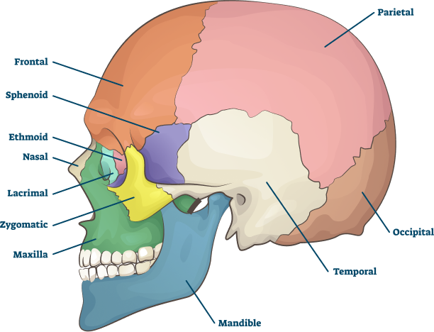
Anatomy Of The Head
| STRUCTURE | DESCRIPTION |
|---|---|
| Skull | Bony structure protecting the brain. |
| Brain | Central organ of the nervous system. |
| Cerebrum | Largest part, involved in thinking and movement. |
| Cerebellum | Coordinates muscle movements, maintains balance. |
| Brainstem | Controls vital functions, connects to spinal cord. |
| Meninges | Three protective layers around brain and spinal cord. |
| Blood Vessels | Arteries and veins supplying and draining blood. |
| Cranial Nerves | Twelve nerves responsible for sensory and motor functions. |
| Ventricles | Cavities filled with cerebrospinal fluid. |
| Sinuses | Air-filled spaces reducing skull weight. |
Alternative Names For CT Head Contrast:
What Is The Purpose Of This Test?
The primary purpose of a CT head contrast scan is to obtain detailed images of the brain and its blood vessels to aid in the diagnosis and evaluation of various neurological conditions. By using a contrast dye, the scan enhances the visibility of abnormalities such as tumours, aneurysms, strokes, and infections, providing a clearer and more precise picture than a standard CT scan. This enhanced imaging is crucial for detecting issues that may not be visible otherwise, allowing for more accurate diagnoses and better-informed treatment plans. Additionally, CT head contrast scans are essential for pre-surgical planning, as they give surgeons a detailed map of the brain’s anatomy, helping to minimise risks during procedures. The test is also used to monitor the progression of certain brain conditions and to assess the effectiveness of ongoing treatments, making it a versatile and indispensable tool in neurological healthcare. Book a CT Head in Raigad for ONLY2000*
CT Head Contrast Available In
Name
Proc. Time
Rating
Price

What Does It Show?
Blood Vessels: Enhanced visualisation of blood vessels, aiding in the identification of aneurysms, arteriovenous malformations (AVMs), and blockages.
Tumours: Detailed imaging of brain tumours, providing information on their size, location, and involvement with surrounding tissues.
Strokes: Clear distinction between ischemic (due to blockage) and hemorrhagic (due to bleeding) strokes.
Infections: Improved detection of infections such as abscesses, meningitis, and encephalitis.
Brain Injuries: Assessment of traumatic brain injuries, including haemorrhages, contusions, and skull fractures.
Structural Abnormalities: Identification of congenital or acquired structural abnormalities within the brain.
Post-Surgical Evaluation: Monitoring post-surgical changes to ensure proper healing and detect complications.
Hydrocephalus: Visualisation of ventricular enlargement indicating cerebrospinal fluid buildup.
Inflammation: Detection of inflammation within brain tissues.
Blood-Brain Barrier Issues: Highlighting areas where the blood-brain barrier is disrupted, indicating potential disease or injury.
Difference Between CT Head Contrast & NCCT Head
When it comes to imaging the brain, two common types of CT scans are often used: CT head contrast and NCCT head (Non-Contrast CT Head). Both provide valuable information, but they serve different purposes and have distinct characteristics. Below is a table outlining the key differences between these two types of CT scans.
| FEATURE | CT HEAD CONTRAST | NCCT HEAD (NON-CONTRAST CT HEAD) |
|---|---|---|
| Definition | A CT scan that uses a contrast dye to enhance image clarity. | A CT scan performed without the use of contrast dye. |
| Purpose | To highlight blood vessels and abnormalities for better visualisation. | To provide a general overview of the brain’s structure. |
| Usage | Used for detailed diagnosis of tumours, aneurysms, strokes, and infections. | Used for initial assessment of head injuries, strokes, and structural abnormalities. |
| Image Clarity | Enhanced clarity due to contrast dye highlighting different tissues. | Standard clarity without enhancement from dye. |
| Diagnosis Capability | Better at detecting and defining the extent of abnormalities. | Sufficient for identifying major issues but less detailed. |
| Procedure | Involves injecting a contrast dye into the bloodstream before scanning. | No injection of dye; straightforward scanning process. |
| Preparation | Requires fasting and possible allergy screening due to contrast dye. | Minimal preparation, usually no fasting required. |
| Risks | Risks include allergic reactions to dye, kidney function impact, and mild side effects like nausea. | Lower risk as no contrast dye is used, but still involves radiation exposure. |
| Radiation Exposure | Similar level of radiation exposure as NCCT, but with added contrast-related risks. | Involves exposure to radiation, though minimal compared to other scans. |
| Cost | Generally more expensive due to the use of contrast dye and additional processing. | Less expensive as it does not involve contrast dye. |
| Typical Applications | Detailed assessment for surgical planning, complex diagnoses, and monitoring treatment efficacy. | Initial evaluation in emergency settings, assessment of acute conditions like trauma and bleeding. |
Health Conditions Diagnosed With A CT Head Contrast
CT head contrast imaging is a valuable tool for diagnosing a wide range of health conditions affecting the brain and its vasculature. Some of the common health conditions diagnosed using CT head contrast include:
Brain Tumours: Contrast-enhanced CT scans help in identifying and characterising brain tumours, including gliomas, meningiomas, pituitary adenomas, and metastatic lesions.
Cerebrovascular Diseases: CT head contrast imaging is essential for diagnosing cerebrovascular conditions, including ischemic and hemorrhagic strokes, cerebral aneurysms, and arteriovenous malformations (AVMs).
Head Trauma: CT scans with contrast are essential for assessing traumatic brain injuries, including intracranial haemorrhages, contusions, and skull fractures.
Intracranial Infections: Contrast-enhanced imaging helps identify brain infections such as abscesses, meningitis, and encephalitis by highlighting areas of inflammation.
Brain Abscesses: Localised collections of pus within the brain parenchyma are detected via CT head contrasts.
Hydrocephalus: CT scans with contrast can visualise enlarged ventricles in cases of hydrocephalus, a condition characterised by the buildup of cerebrospinal fluid in the brain.
Intracranial Hematomas: Contrast-enhanced imaging helps differentiate various types of intracranial hematomas, such as epidural, subdural, and intraparenchymal hematomas.
Brain Metastases: Contrast-enhanced CT scans are useful for detecting and monitoring metastatic lesions in the brain, particularly in patients with known primary cancers.
Vascular Lesions: CT head contrast can identify other vascular lesions such as cavernous malformations, venous thrombosis, and vasculitis.
Intracranial Masses: CT head contrast aids in the characterisation of various intracranial masses, including cysts, hematomas, and inflammatory pseudotumours.
These are just a few examples of the many health conditions diagnosed using CT head contrast imaging. The technique is versatile and plays a crucial role in the diagnosis, treatment planning, and monitoring of neurological disorders. Book a CT Head in Raigad for ONLY2000*
Who Should Get Tested?
Individuals With Neurological Symptoms: Individuals experiencing symptoms such as sudden weakness, numbness, difficulty speaking, or visual disturbances may require a CT head contrast scan to investigate potential underlying neurological conditions.
Patients With Head Trauma: Patients who have suffered significant head injuries, such as from accidents or falls, may undergo a CT head contrast scan to evaluate for traumatic brain injuries, including intracranial haemorrhages and skull fractures.
Individuals With Severe Headaches: Individuals experiencing persistent or severe headaches that do not respond to standard treatments may benefit from a CT head contrast scan to investigate potential underlying causes such as tumours or vascular abnormalities.
Patients With Suspected Brain Abnormalities: Patients presenting with symptoms or clinical findings suggestive of brain abnormalities may undergo imaging with CT head contrast to obtain detailed anatomical information and aid in diagnosis.
Individuals With Neurological Conditions: Individuals with known neurological conditions or a history of strokes, aneurysms, or brain tumours may undergo periodic CT head contrast scans to monitor disease progression, treatment response, or recurrence.
Patients Undergoing Pre-Surgical Evaluation: Patients scheduled for neurosurgical procedures or interventions may require a CT head contrast scan as part of pre-operative assessments to identify pre-existing conditions or anatomical abnormalities.
Individuals With Altered Mental Status: Individuals presenting with altered mental status, confusion, or other cognitive impairments may undergo a CT head contrast scan to assess for potential causes such as brain infections, haemorrhages, or tumours.
Patients With Acute Neurological Symptoms: Patients experiencing acute neurological symptoms such as seizures, loss of consciousness, or focal neurological deficits may undergo urgent CT head contrast imaging to rule out life-threatening conditions such as intracranial bleeding or tumours.
Ultimately, the decision to undergo a CT head contrast scan should be made in consultation with a healthcare provider, who will consider the individual’s symptoms, medical history, and clinical presentation before recommending the appropriate imaging study. Book a CT Head in Raigad for ONLY2000*
How Does CT Head Contrast Work?
CT head contrast imaging works by combining the principles of computed tomography (CT) scanning with the use of a contrast agent to enhance the visibility of structures within the brain. Here’s how it works:
Injection Of Contrast Agent: Before the scan, a contrast agent containing iodine is injected intravenously into the patient’s blood. This agent circulates through the body and reaches the blood vessels within the brain.
Scanning Process: The patient lies on a table that moves into the CT scanner, which consists of an x-ray tube and detectors. The x-ray tube rotates around the patient, emitting x-ray beams that pass through the body.
X-Ray Absorption: As the x-ray beams pass through the body, they are absorbed to varying degrees by different tissues. Dense tissues, such as bone, absorb more x-rays and appear white on the resulting images, while less dense tissues, such as soft tissue and fluids, allow more x-rays to pass through and appear darker.
Contrast Enhancement: In areas where the iodine-based contrast agent has accumulated, such as blood vessels within the brain, the x-ray absorption is increased. This results in enhanced contrast between the blood vessels and surrounding tissues on the CT images.
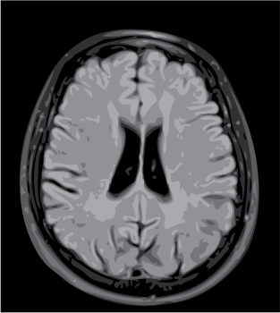
Image Reconstruction: The detectors in the CT scanner capture the x-rays that pass through the body from multiple angles. This data is processed by a computer to reconstruct cross-sectional images/slices of the brain.
Visualisation: The reconstructed CT images are displayed on a monitor, allowing radiologists and physicians to visualise the internal structures of the brain. The contrast-enhanced images provide clearer delineation of blood vessels, tumours, areas of bleeding, and other abnormalities compared to non-contrast CT scans.
Overall, CT head contrast imaging enables healthcare providers to obtain detailed and precise information about the structures and conditions within the brain, aiding the diagnosis and management of neurological disorders. Book a CT Head in Raigad for ONLY2000*
CT Head Contrast Procedure
Patient Preparation: Patients are instructed to remove metal objects that may interfere with imaging. Prior medical history, including allergies, is carefully reviewed to ensure patient safety.
Contrast Injection: A contrast agent containing iodine is administered intravenously to enhance visualisation of blood vessels and the brain. This is performed by a trained professional using a syringe/automated injector.
Patient Positioning: Patients are positioned on the CT scanner table, usually lying flat on their back with their head secured in place to minimise motion artefacts during imaging.
CT Scan Acquisition: The CT scanner rotates around the patient's head, emitting x-ray beams that pass through the skull and brain tissues. Multiple cross-sectional images of the brain are acquired at various angles.
Image Acquisition: Detectors measure the intensity of transmitted radiation. The contrast agent enhances the visibility of blood vessels, tumours, and other abnormalities, providing detailed images for analysis.
Monitoring: Patients are closely monitored throughout the procedure, with continuous observation by a radiologist or healthcare provider. Any discomfort or adverse reactions to the contrast agent are addressed.
Post-Scan Observation: After the CT scan, patients may be observed for a brief period to monitor for any immediate adverse reactions, such as allergic responses or contrast-induced nephropathy.
Image Interpretation: The acquired CT images are interpreted by a radiologist, who checks for tumours, haemorrhages, or vascular lesions. Findings are captured in a comprehensive report for further management.
The duration of a CT head contrast typically varies depending on factors such as the protocol used, the complexity of the case, and any unforeseen circumstances during the procedure. On average, the actual scanning time ranges from a few minutes to around 10-20 minutes. However, this does not account for the additional time required for patient preparation, contrast injection, positioning on the CT scanner table, and post-scan observation. Overall, patients can expect
| MRI Scans | City | Price | ||
|---|---|---|---|---|
| 1 | CT Head Contrast | - | 2000 |
NORMAL TEST RESUTS
Absence Of Abnormalities: A normal result from a CT head contrast scan indicates the absence of significant abnormalities or pathological findings within the brain, blood vessels, and surrounding structures.
Normal Brain Anatomy: It suggests the brain structures like the cerebral cortex, white matter, basal ganglia, and brainstem appear within expected parameters without evidence of lesions, masses, or abnormal density.
Intact Blood Vessels: Normal results indicate that the blood vessels within the brain, including the arteries and veins, exhibit normal morphology and patency, without evidence of stenosis, aneurysms, or thrombosis.
No Evidence Of Hemorrhage: Normal findings suggest the absence of intracranial hemorrhage, such as subarachnoid hemorrhage, intracerebral hemorrhage, or epidural hematoma, which are indicative of acute injury or vascular pathology.
No Acute Pathological Processes: It implies that there are no acute pathological processes, such as acute infarction, inflammatory changes, or infectious processes, affecting the brain or surrounding tissues.
Limited Scope: However, it’s important to note that a normal result on a CT head contrast scan does not rule out all potential abnormalities or underlying conditions. Some conditions may not be detectable by CT imaging alone, and further evaluation may be warranted based on clinical symptoms, history, or other diagnostic tests.
Baseline For Comparison: A normal result provides a baseline for comparison in future imaging studies and helps establish the absence of significant pathology at the time of the scan.
Clinical Correlation: The interpretation of a normal result should be correlated with the patient’s clinical presentation, medical history, and other diagnostic findings to ensure a comprehensive assessment.
ABNORMAL TEST RESULTS
Presence Of Pathology: An abnormal result from a CT head contrast scan indicates the presence of pathological findings or abnormalities within the brain, blood vessels, or surrounding structures.
Identifies Specific Abnormalities: It may reveal various abnormalities such as tumours, cysts, haemorrhages, infarctions, abscesses, or vascular lesions, which may require further evaluation and management.
Diagnostic Significance: Abnormal findings on a CT head contrast scan can provide diagnostic information, guiding clinicians in determining the cause of symptoms and planning appropriate treatment strategies.
Clinical Implications: The presence of abnormalities may have significant clinical implications, depending on the nature and severity of the pathology. For example, the identification of a brain tumour may necessitate surgical intervention, radiation therapy, or chemotherapy.
Potential Prognostic Indicators: Abnormal results may also serve as prognostic indicators, helping clinicians assess the severity of the condition and predict the patient’s prognosis.
Monitoring And Follow-up: Detection of abnormalities may warrant ongoing monitoring and follow-up imaging studies to track disease progression, response to treatment, or the development of complications.
Patient Counselling And Management: Abnormal results require clear communication with the patient regarding the findings, potential implications, and recommended management strategies. This may involve referral to specialists, initiation of treatment, or further diagnostic investigations.
Multidisciplinary Approach: Management of abnormal results often involves a multidisciplinary approach, with collaboration between radiologists, neurologists, neurosurgeons, oncologists, and other healthcare professionals to ensure comprehensive care for the patient.
In summary, an abnormal result from a CT head contrast scan signifies the presence of pathological findings or abnormalities within the brain and surrounding structures. These findings have significant diagnostic and clinical implications, guiding further evaluation, treatment planning, and ongoing management. Effective communication and collaboration among healthcare providers are essential to ensure timely and comprehensive care for patients with abnormal CT scan results.Book a CT Head in Raigad for ONLY2000*
Associated Risks
Allergic Reactions: Some patients may experience allergic reactions to the iodine-based contrast agent, ranging from mild itching or rash to severe anaphylactic reactions. It's crucial to inform the healthcare provider of any known allergies.
Kidney Damage: The contrast agent can potentially cause kidney damage, particularly in patients with pre-existing kidney disease or those at high risk for renal impairment. Monitoring kidney function before and after the procedure is important.
Radiation Exposure: CT scans involve exposure to ionising radiation, which carries a small risk of developing cancer over a lifetime, particularly with repeated exposure. The benefits of accurate diagnosis generally outweigh this risk.
Injection Site Issues: Complications at the injection site, such as pain, swelling, or infection, can occur. Careful technique and monitoring reduce these risks.
Contrast-Induced Reactions: Some patients may experience nausea, vomiting, or a metallic taste in their mouth after the contrast injection. These reactions are usually mild and temporary.
Rare Complications: In rare cases, the use of contrast agents can lead to more severe complications, such as contrast-induced nephropathy or thyroid dysfunction. These risks are generally low and managed by careful patient selection and monitoring.