CT Scan Vs MRI: Differences And Which One To Go For
Overview of CT Scan vs MRI
Doctors often prescribe scans in order to understand the exact nature of the issue that a person is suffering from. There are many types of scans—with X-ray still being the commonly known name—that aid doctors in not only diagnosing diseases but also in monitoring current conditions. Another two terms that are widely used in the patient world are MRI and CT scan. While they both perform scanning of the human body, the knowledge pertaining to the differences between MRI and CT scan and which scan will be required under what circumstances is still limited.
MRI
MRI or Magnetic Resonance Imaging is a scanning technique that uses magnetic fields along with pulses of radio frequency and a computer to get images of the internal organs, soft tissue, and bone.
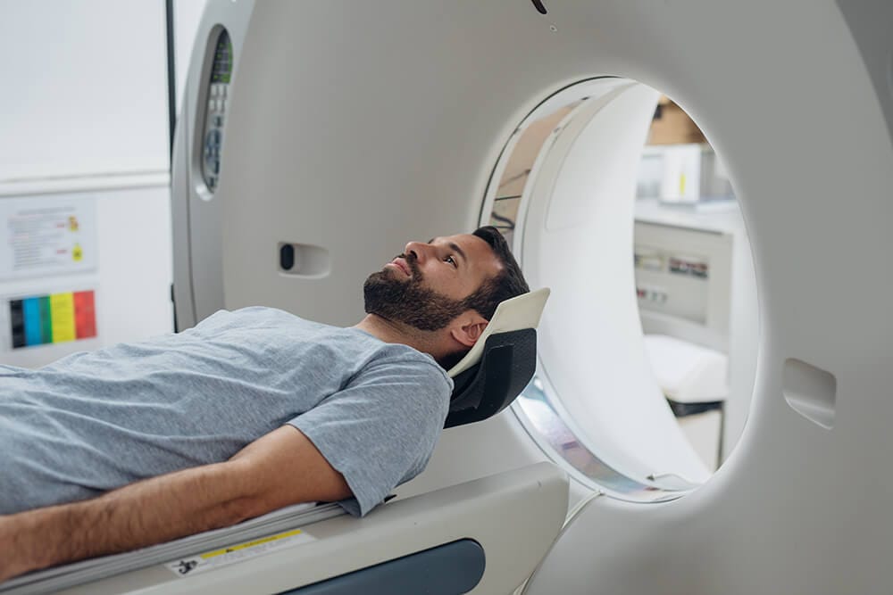
A patient who is suggested to get an MRI is made to lie on a table and then slide within a circular machine. The radio pulses are collected via a coil that acts as an antenna, and the images are produced on the computer screen. This coil is placed on the part of the body that requires scanning. MRI is used to get:
-
- Images of internal organs and the structures of bones
- Difference between normal tissues and abnormal tissues
- Radiation-free images
CT scan
CT or Computerized Tomography scan is another method of getting X-rays of certain body parts and internal structures via radiation. The required cross-sectional images of the body parts are generated by laser thin beams by scanning. The images are produced on the computer linked to the machine.
This is the most common type of scanning technique, after the X-ray, to get a detailed understanding of the issues faced by patients, especially those who have cancer. People with metal bits in them can opt for a CT scan, unlike MRI, due to the absence of magnetic fields in the CT technique. A CT scan is helpful for getting:
-
- Images of blood vessels, along with bone and tissue structures
- A detailed view of joint injuries
- An understanding of the issues related to chest pain
Injuries that require the use of CT scan or MRI
Both MRI and CT scan are used to scan the internal structure of a person’s body. While scanning can reflect various issues, both these techniques have their own set of usages in terms of injury scanning. In order to solve the dilemma of choosing CT scan vs MRI for scanning the brain, understanding the range of both in terms of scanning prowess is required.
MRI is used for checking and diagnosing:
-
- Injuries or abnormalities in the joints and ligaments.
- Breast cancer.
- Tumors.
- Irregularities related to blood vessels, like detection of aneurysms, artery blockages, or injuries sustained from a previous case of myocardial infarction.
- Problems related to the brain, like diagnosing multiple sclerosis, stroke, or aneurysms.
- Understanding the root cause of the injuries that affect the spinal cord.
- Liver disorders, like cirrhosis.
- Bowel conditions caused by Crohn’s disease or ulcerative colitis.
- Disorders related to the bones, such as osteonecrosis, bone marrow disease, and herniation, or degeneration caused to discs and spine.
CT scan is used for checking and diagnosing:
-
- Issues related to the circulatory system and general circulation, like heart diseases, blockages of blood vessels, aortic aneurysms, or pulmonary edema.
- Kidney disorders, like polycystic kidney disease, or detection of tumors in kidneys.
- Issues and abnormalities related to the abdomen, like unnatural masses around organs.
- Causes for the presence of blood in the urine.
- Diseases related to the lungs, like emphysema, tumors, fibrosis, pleural effusion, or collapsed lung.
- Further proof of skeletal damages that are not often detected by the X-ray, such as osteoporosis or spinal cord injuries.
- Injuries to the head, like brain calcification, hemorrhage, or issues related to blocked blood flow to the brain.
Benefits of using CT scan and MRI
The MRI and CT scan are mainly used to detect the presence of cancer and to check the progression of cancerous cells, when cancer has already been diagnosed and treatment is ongoing. These scanning devices trump traditional X-ray devices, as the former are more detailed in terms of in-depth diagnosis. The techniques differ but the end result remains very similar. Both these scanning methods have their own usages and advantages.
Benefits of MRI:
-
- MRI uses a method that does not pose any risk of being exposed to ionizing radiation.
- MRI also poses no risk of contracting infection via the method or while examination is ongoing.
- There is no scope of developing deep tissue burns with an MRI that are often the case with scans using ionizing radiation.
- The process of MRI is non-invasive and does not make patients uneasy about privacy or other health concerns.
- MRI helps with getting a wider range and sectional view of the patient’s body when compared to an ordinary X-ray scan. MRI also provides more detailed images of the soft tissues and helps determine abnormal tissues.
CT scans also check for the same injuries and abnormalities that are examined via an MRI. Though CT scan uses ionizing radiation, there are many advantages that it has over an MRI.
Benefits of a CT scan:
-
- As compared to an MRI, CT scans are less expensive and less noisy.
- CT scans are also very comfortable as the examination happens within minutes. MRIs, on the other hand, can take somewhere between 10 minutes to even an hour.
- CT scan machines are open in terms of the design of the machine. This also helps reduce anxiety in patients.
Procedure of using CT scan and MRI
The procedure for scanning using the CT scan and the MRI are similar in some ways due to the use of a circular device to scan the whole body while the patient lies down on a bed/tunnel.
The MRI scan uses a superconducting magnet that sends radiofrequency waves within the body. The atoms are arranged in either the north or south position and a few are left unmatched. When radiofrequency is on, these unmatched atoms start spinning in opposite directions. When the frequency is turned off, these atoms return to their original position and emit an energy that is then recorded and converted to images seen on a screen.
The CT scan takes multiple X-ray images from various angles which are then used to construct a 3-dimensional image of the organ that is being diagnosed. Sometimes, a contrast material, such as a dye, can be used on a patient while doing a CT scan. This contrast material is used to block certain areas to help highlight other areas.
Risks of using CT scan and MRI
In a CT scan, the body goes through radiation. On the other hand, in an MRI scan, the body is subjected to a magnetic field. Both these scanning techniques offer in-depth details about the diagnosis and help doctors understand the ailments of a patient. But, there are certain risks that are associated with both these methods.
MRI does not use ionizing radiation, but since a magnetic field is introduced, metal implants cannot be introduced to the MRI. People having implants like pacemakers, cochlear, intracranial aneurysm clips, prosthetic devices, IUDs, stimulators for bone growth, neurostimulators, or drug infusion pumps should not opt for MRI. Pregnant women are also advised to not go for an MRI, even if they are required to get a deep scanning done.
CT scan is safe for people with metallic implants, but since ionizing radiation is used, chances of damage occurring to the DNA or even cancerous growth can be possible. Contrast material can cause allergic reactions in some patients. Thus, care has to be taken. Pregnant women should not be introduced to the scan, as damage to the fetus is possible.
Choosing between CT scan and MRI
Both CT scans and MRIs are used to check for internal damages or disorders in internal organs. But, in order to understand which scan technology to opt for, the kind of injury and the suggestions of the doctor have to be taken into consideration. Generally, a CT scan is suggested when a general idea about the internal organs or an estimate of any trauma induced by head injury or fracture is required. The MRI is suggested for a more detailed diagnosis:
-
- Injuries sustained by the soft tissues
- Herniated disks
- Ligament damage like lorn ligament
A quick comparison of CT scan vs MRI
CT Scan |
MRI |
| Is more cost-effective | Is more expensive |
| Produces less sound | Produces more sound |
| A CT scan is faster, takes less than 5 minutes | MRI is time-consuming and can even last for one hour |
| Images are not very clear | Images produced are very sharp and precise |
| Utilizes ionizing radiation | No ionizing radiation used for scanning |
| Ideal for people who are built larger | Not ideal for large built people |
| Metallic implants do not affect scans | People with metallic implants cannot opt for an MRI |
| Contrast material like dyes are used that can cause allergic reactions | No such reactive materials are used |
Precautions to be taken for MRI and CT scan
As mentioned above, there are certain risks associated with both these scanning methods. Therefore, precautions should be taken. If a woman is pregnant or pregnancy is suspected, before opting for either of these scans, an ultrasound should be conducted first. The MRI should be avoided if the person has any metallic implants.
Conclusion
MRI and CT scans help detect deeper issues and cancerous cells. People who are suspected to have cancer or have sustained an injury to the head and suspect a concussion can go for these scans which help solve various diseases. With proper precaution, these scans can be carried out with ease.
FAQs
What is an Aneurysm clip?
The Aneurysm clip is a microsurgical implant that is used to stop blood flow into the aneurysm.
Are CT scans and MRI painful?
These scanning techniques are non-invasive and do not require any special preparation on the part of the patient. But, the techniques require the person to lie down for some time, which might cause some discomfort, especially during an MRI.
What is meant by soft tissues?
Soft tissues are generally muscles, tendons, ligaments, body fat, or fascia. These tissues provide support to the bones.
References


